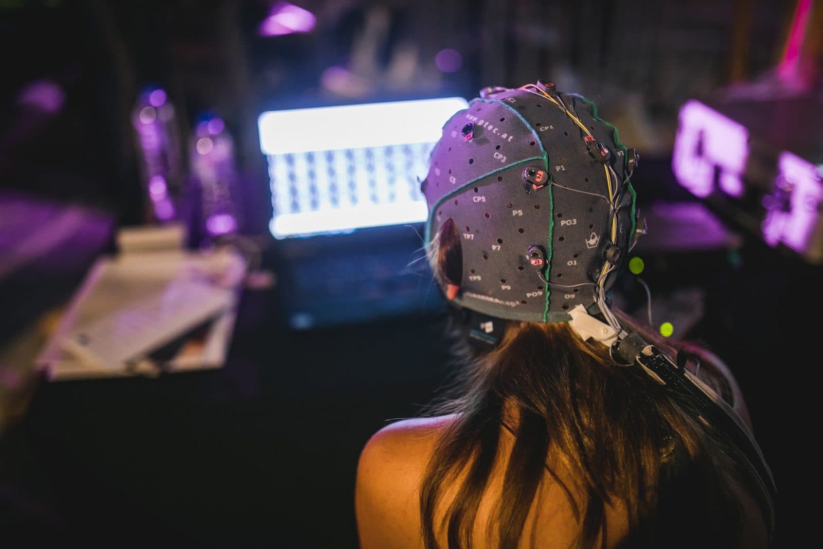
What is tDCS? Transcranial direct current stimulation
What is tDCS: how transcranial direct current stimulation is used in research; electrodes and montages, current and duration, sham, EEG and safety.
Published
Transcranial direct current stimulation (tDCS) is a non-invasive brain stimulation method that temporarily changes the excitability of the cortex by passing a weak, constant direct current between two or more electrodes placed on the scalp. This article treats tDCS as a research method: how it works, how montages and dose parameters are chosen and reported, sham control, combining it with EEG, and the safety guidelines. Clinical use requires a physician’s assessment and is not the subject of this article.
How does tDCS work? Anodal and cathodal stimulation
tDCS does not fire neurons directly. The weak electric field the current creates in the brain slightly shifts the resting membrane potential of neurons and so changes the probability that they fire. For this reason tDCS is usually described not as a stimulator in the strict sense but as a method that modulates cortical excitability. The current enters through the electrode called the anode and leaves through the cathode; the effect under the two electrodes can go in opposite directions.
In their landmark study, Nitsche and Paulus (2000) measured the effect of weak direct current over the motor cortex with TMS and showed that anodal stimulation increased excitability while cathodal stimulation decreased it. The change reached up to 40 per cent, lasted several minutes after the end of stimulation, and its strength and duration varied with the current intensity and the stimulation duration. This finding is the basis of the polarity principle often summarised as “excitation under the anode, inhibition under the cathode”.
The relationship is not always that simple, however. Batsikadze et al. (2013) reported that cathodal stimulation at 2 mA for 20 minutes over the motor cortex increased excitability instead of decreasing it, whereas cathodal stimulation at 1 mA produced the expected decrease. Because the effect can vary with current intensity, duration, target region and the individual, the polarity effect should be treated not as an assumption but as a hypothesis to be tested in each study.
Electrodes and montages
A classic tDCS set-up has two electrodes: the anode and the cathode. Woods et al. (2016) describe the most common electrode assembly as a metal or conductive rubber electrode, a sponge around it, and an electrolyte (saline, gel or conductive cream) that eases the passage of current into the skin. The sponge should be soaked enough but not too much: saline overflowing from an over-saturated sponge spreads over the scalp outside the electrode, enlarges the area through which current passes and undermines the reproducibility of the application. Electrode sites are usually defined by positions of the EEG 10-20 system, which allows a montage to be repeated in other laboratories.
Some of the montages often met in the literature are:
- Motor cortex montage
- The anode is placed over the primary motor cortex (around C3 or C4) and the cathode over the opposite eyebrow (the supraorbital region). It was used in Nitsche and Paulus’s studies and is one of the most studied arrangements in the tDCS literature.
- Dorsolateral prefrontal cortex (DLPFC) montage
- The anode is placed around F3 or F4 and the cathode on the opposite side or over the supraorbital region; it is often used in studies of cognitive processes such as working memory.
- Extracephalic reference
- The return electrode is placed off the head, for example on the shoulder or arm, to reduce the effect of the second electrode on the brain. Woods et al. (2016) note that this arrangement may require a higher stimulation intensity.
- High-definition (HD) tDCS
- A small central electrode over the target is surrounded by four return electrodes (a 4×1 ring), concentrating the current in a smaller area.
Whichever montage is chosen, the current passes through a large part of the brain between the two electrodes; the target is not only the area under the anode. Woods et al. (2016) stress that physiological measurements and computer models predicting current flow show that the relative position of the electrodes markedly changes where, and how much, current reaches the brain. It is therefore good practice to estimate the electric field with models when planning a montage, and to report electrode positions, sizes and the location of the return electrode in detail in the methods section.
Current, current density and duration
The main parameters that define a tDCS dose are current intensity, electrode size, stimulation duration and montage. Woods et al. (2016) give 1–2 mA as typical. A frequently used arrangement in motor cortex studies is 1 mA delivered through 35 cm² (5×7 cm) electrodes (Batsikadze et al., 2013); with this arrangement, after-effects lasting about an hour have been reported after 9 minutes of stimulation under the cathode and 13 minutes under the anode (Woods et al., 2016). These are typical values from the literature, not recommendations: each study’s protocol is set with ethics committee approval and on the basis of published guidelines.
| Parameter | Definition | Why it is reported |
|---|---|---|
| Current intensity (mA) | The constant current passing between the electrodes | Determines the size of the electric field in the brain |
| Electrode type and size | Sponge, gel or HD electrode; surface area (cm²) | Affects current density and distribution under the skin |
| Current density | Current divided by electrode area (mA/cm²) | Affects skin sensations and the safety assessment |
| Duration | Stimulation time, including the ramps that switch the current on and off | Relates to the size and persistence of the effect |
| Montage | Position of anode and cathode by their 10-20 names | Determines which regions the current passes through |
| Number and spacing of sessions | A single session or repeated sessions | Important for cumulative effects and safety monitoring |
| Total charge | Current × time (coulombs) | A dose measure used in the safety literature |
A simple calculation makes doses easier to compare: 1 mA through a 35 cm² electrode gives an average density of about 0.029 mA/cm², and a 20-minute session at 2 mA delivers a total charge of 2.4 coulombs. Switching the current on and off with ramps lasting a few seconds is common; Woods et al. (2016) note that the ramps prevent the perception of phosphenes (flashes of light) that an abrupt onset or offset can cause.
Sham stimulation and blinding
Most tDCS studies use sham stimulation to separate the effect from placebo and expectation. Woods et al. (2016) describe the usual method: the current is ramped up and down as in real stimulation but held at the target intensity for only a few seconds. The participant feels the initial itching or tingling, but no current is delivered for the rest of the session. Gandiga et al. (2006) reported that tDCS and sham stimulation over the motor cortex caused comparably minimal discomfort and that neither the participants nor the investigators questioning them could tell the two conditions apart.
Even so, the success of blinding should be measured in each study, not assumed. Skin sensations can become more noticeable at higher currents, and participants who have been stimulated before may guess the condition. Many protocols therefore ask participants at the end of the session which condition they think they received, and prefer double-blind designs in which the person applying stimulation does not know the condition either; some research devices offer a coded study mode for this purpose. Antal et al. (2017) note that adverse effects are frequently reported by participants receiving placebo stimulation too, which means the sham group’s adverse effects should be recorded at least as carefully as those of the real stimulation group.
tDCS and EEG together
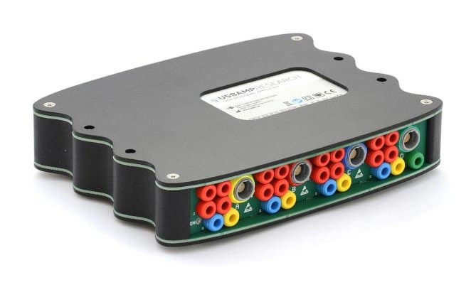
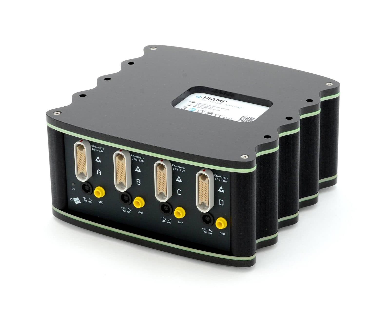
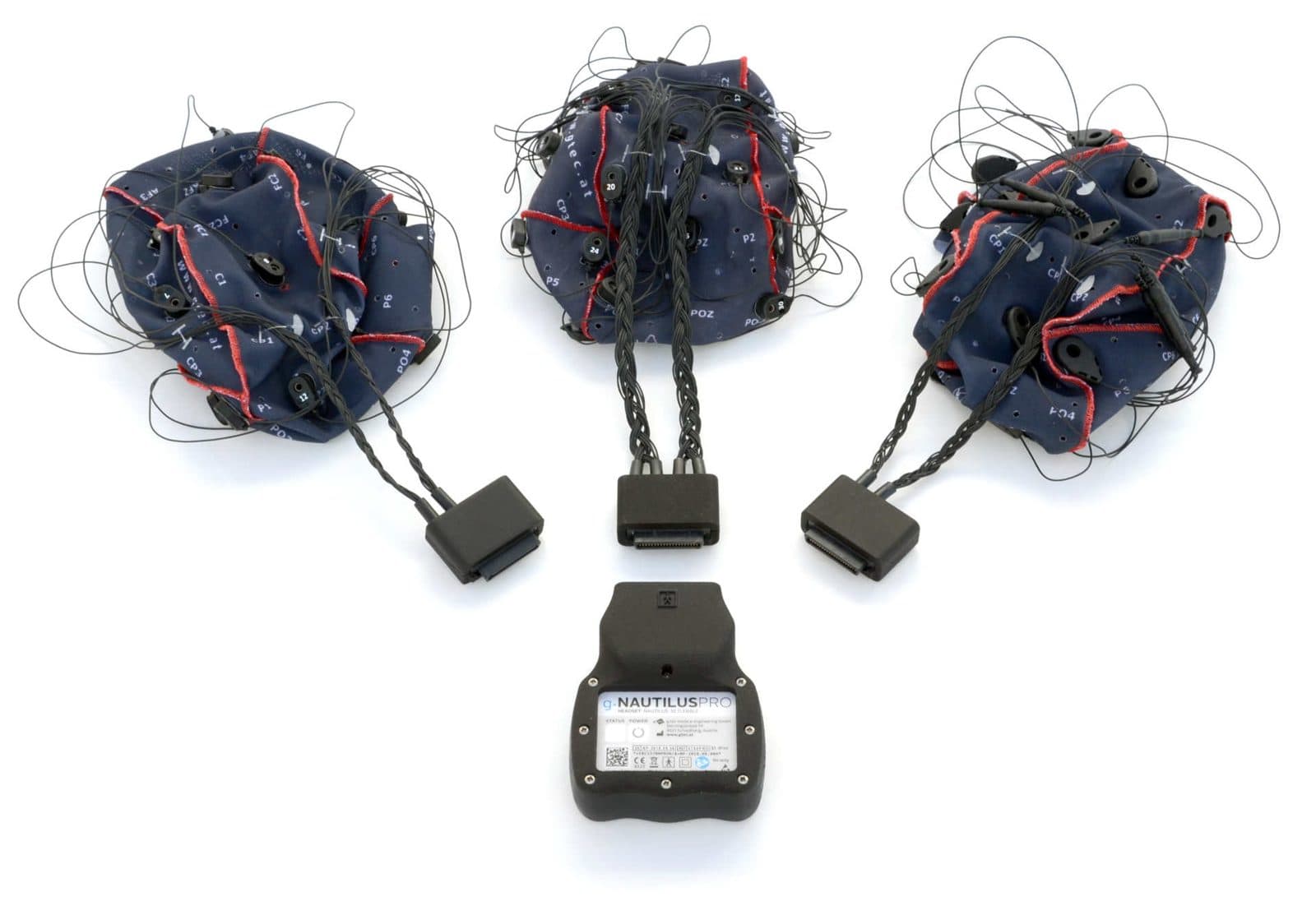
Combining tDCS with EEG to see how it changes brain activity is increasingly common. There are two arrangements: separate EEG recordings before and after stimulation, or EEG recorded while stimulation is running. In simultaneous recording, the applied direct current and especially the changes during the ramps can cause large shifts and artefacts in the EEG. This calls for an amplifier with a wide input range that is DC-coupled and is not driven into saturation by the stimulation current, and for analysis tools that remove these artefacts. Antal et al. (2017) report that accompanying procedures such as electrophysiological measurements were not associated with additional safety issues.
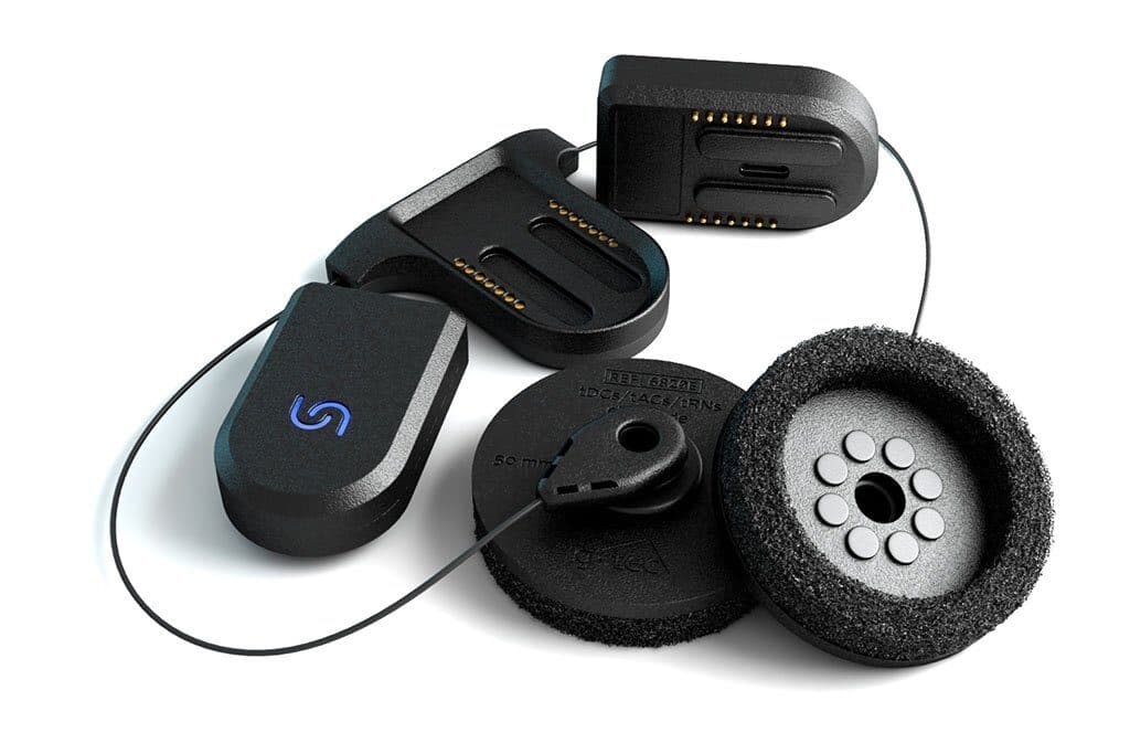
In the catalogue, g.USBamp Research is described as suited to EEG-tDCS recordings thanks to its DC-coupled design and ±250 mV input range; g.HIamp and g.Nautilus Pro Flexible also support EEG recording simultaneously with TMS and tDCS. According to the manufacturer, g.BSanalyze includes dedicated algorithms for removing TMS and tDCS stimulation artefacts. Electrode placement should be planned jointly as well: since tDCS electrodes overlap positions on the EEG cap, which EEG channels will be recorded is designed together with the montage. The catalogue’s Unicorn tDCS Core-2 is a wearable tDCS device controlled from a smartphone, with one cathode and one anode and a current adjustable from 0 to 2 mA.
Safety and ethics
The most comprehensive consensus document for low-intensity transcranial electrical stimulation is the safety, ethical, legal regulatory and application guideline published by Antal et al. (2017) in Clinical Neurophysiology. The guideline summarises that no serious adverse events have been reported in over 18,000 sessions given to healthy participants and to neurological and psychiatric patients. Moderate adverse events are rare; in tDCS the main one is skin burns caused by poor contact between electrode and skin. Mild adverse events include headache and fatigue after stimulation and prickling and burning sensations during it. The guideline states that safety has been established for “conventional” applications below 4 mA lasting up to 60 minutes a day; it also notes that mania or hypomania was very rarely induced in patients with depression (11 documented cases), while a causal relationship is difficult to prove.
Bikson et al. (2016) likewise report that human studies with conventional protocols (at most 40 minutes, at most 4 mA, at most 7.2 coulombs) produced no report of a serious adverse effect or irreversible injury across more than 33,200 sessions. In animal studies, brain injury occurred at levels more than an order of magnitude above the estimated current densities produced by conventional tDCS. These figures are not a guarantee of safety but a summary of the literature to date. In practice, research protocols rest on these principles:
- The study is run with ethics committee approval and informed consent; exclusion criteria (for example intracranial metal implants, a history of seizures, skin lesions at the electrode site) are defined in the protocol.
- Contact between electrode and skin is checked in every session; electrodes are not placed over damaged skin.
- Adverse effects are recorded in every session with a structured questionnaire; Antal et al. (2017) propose structured interview forms for this purpose.
- The device is used only by trained researchers and with the parameters set in the protocol.
Redness under the electrodes is usually due to vasodilation, and Woods et al. (2016) do not consider it a safety issue on its own; it should still be recorded and distinguished from a burn. Clinical use is a separate matter: it requires a physician’s assessment, an appropriate indication and devices approved for that purpose. This article contains no diagnostic or treatment advice.
How tDCS differs from TMS
Although tDCS and transcranial magnetic stimulation (TMS) are both listed under non-invasive brain stimulation, their physical principles, hardware and roles in research differ:
| Feature | tDCS | TMS |
|---|---|---|
| Form of stimulation | A weak, constant direct current between electrodes | Brief, strong magnetic pulses from a coil that induce current in the cortex |
| Effect on neurons | Shifts the membrane potential and changes firing probability; does not trigger action potentials directly | At sufficient intensity, triggers action potentials directly (for example a motor evoked potential) |
| Focality | Relatively diffuse; more focal with HD montages | Relatively focal; depends on the coil type |
| Time structure | Continuous stimulation lasting minutes | Single pulses or repetitive pulse trains |
| Participant experience | Tingling, itching, a mild burning sensation | A clicking sound, twitching sensations in the scalp and muscles |
| Sham method | Current ramped on and switched off shortly after | A sham coil or a changed coil angle; matching the sound and touch sensation is planned separately |
| Hardware | Small, battery-powered, portable stimulators | Larger stimulators and coils; usually a fixed installation in a lab or clinic |
| Together with EEG | Continuous current and ramp artefacts; needs a DC-coupled amplifier with a wide input range | A brief, very large pulse artefact; needs a TMS-compatible amplifier and electrodes |
In short, TMS delivers a stimulus and lets you measure the cortex’s response, while tDCS shifts the excitability of the cortex for a period of time. That is why the two methods are often used together: as in Nitsche and Paulus’s (2000) study, the effect of tDCS is assessed through changes in motor evoked potentials measured with TMS. You will find TMS covered in detail in the knowledge bank’s TMS article. For information and a quote on the catalogue’s tDCS and stimulation devices and EEG amplifiers, you can use the quote form.
Frequently asked questions
What is tDCS?
Transcranial direct current stimulation (tDCS) is a non-invasive brain stimulation method that temporarily changes the excitability of the cortex by passing a weak, constant direct current between electrodes placed on the scalp. It does not fire neurons directly; it shifts their membrane potential and so changes the probability that they fire. In research it is used to study motor, cognitive and emotional processes.
What is the difference between anodal and cathodal tDCS?
Foundational studies showed that anodal stimulation over the motor cortex increases excitability and cathodal stimulation decreases it. The effect can, however, vary with current intensity, duration, target region and the individual; for example, cathodal stimulation at 2 mA for 20 minutes has been reported to increase excitability. The polarity effect is therefore a hypothesis to be tested in each study.
What is sham tDCS?
Sham tDCS is a placebo stimulation that imitates the initial sensation of real stimulation: the current is ramped up and down and held at the target intensity for only a few seconds. The participant feels the initial tingling, but no current is delivered for the rest of the session. The aim is to separate the effect from expectation and placebo; the success of blinding should be measured by asking participants at the end of the session.
How is a tDCS montage chosen?
The montage is chosen according to the target region in the research question and the path the current will take through the brain. Electrode positions are usually defined by the EEG 10-20 system; for the motor cortex, an anode over C3 or C4 with the cathode over the opposite supraorbital region is a common arrangement. Because the current passes through a large area between the two electrodes, estimating the electric field with computer models is good practice.
Is tDCS safe?
The consensus guideline by Antal et al. (2017) states that no serious adverse events have been reported in over 18,000 sessions and that safety has been established for conventional applications below 4 mA lasting up to 60 minutes a day. The most common adverse effects are tingling, a burning sensation, headache and fatigue; poor electrode contact can cause skin burns. Research is carried out with ethics approval, exclusion criteria and adverse effect monitoring, while clinical use requires a physician’s assessment.
What is the difference between tDCS and TMS?
TMS induces current in the cortex with brief magnetic pulses from a coil and, at sufficient intensity, can fire neurons directly. tDCS passes a weak, constant current between electrodes and changes the excitability of neurons over minutes, but does not trigger action potentials directly. The two methods are often used together in research; the effect of tDCS can be measured with TMS.
References and further reading
- Antal et al. (2017) · Low intensity transcranial electric stimulation: safety, ethical, legal regulatory and application guidelines, Clinical Neurophysiology
- Bikson et al. (2016) · Safety of transcranial direct current stimulation: evidence based update 2016, Brain Stimulation
- Woods et al. (2016) · A technical guide to tDCS, and related non-invasive brain stimulation tools, Clinical Neurophysiology
- Nitsche and Paulus (2000) · Excitability changes induced in the human motor cortex by weak transcranial direct current stimulation, The Journal of Physiology
- Batsikadze et al. (2013) · Partially non-linear stimulation intensity-dependent effects of direct current stimulation on motor cortex excitability in humans, The Journal of Physiology
- Gandiga, Hummel and Cohen (2006) · Transcranial DC stimulation (tDCS): a tool for double-blind sham-controlled clinical studies in brain stimulation, Clinical Neurophysiology