What is neuromarketing? Methods and setting up a lab
What is neuromarketing (consumer neuroscience)? What EEG, eye tracking, EDA and fNIRS measure, their limits and ethics, and how to set up a neuromarketing lab.
Published
What is neuromarketing?
Neuromarketing is the research field that studies how consumers respond to products, brands, advertisements and prices through measurements of the brain and body; in academia it is more often called consumer neuroscience. Its methods are those of cognitive neuroscience and psychophysiology: EEG, eye tracking, electrodermal activity (EDA), heart rate, fNIRS, facial expression analysis and, in academic studies, fMRI.
A distinction often made in the literature is that consumer neuroscience refers to academic research aimed at understanding the processes behind decision making and consumer behaviour, while neuromarketing refers to applying that knowledge and those methods in practice, for example in advertising, packaging and product tests. Plassmann et al. (2015) note that the field’s first decade mainly established the basic neural processes of judgement and decision making, and that for the field to thrive this work must be carried into marketing theory and practice. Either way, the aim is to see not only what participants say but also the reactions they cannot put into words or do not notice: the second of an advertisement at which attention drifts, the product on the shelf that is never seen, the step of an interface at which mental load rises.
What does neuromarketing research study?
The subject of neuromarketing studies is every moment at which a person meets a marketing stimulus. Common questions include the following:
- Advertising and video: in which scene attention gathers or drifts, and when the brand and product are seen.
- Packaging and shelf: whether a product is noticed among its competitors, and in what order the information on the label is read.
- Websites and apps (UX): the path users take until they find what they are looking for, the mental load at moments of decision, and the steps where errors occur.
- Price and choice: when and how closely price information is examined, and how gaze moves while choosing between options.
- Brand and experience: body and brain responses during real experiences such as tasting, a shop’s atmosphere or driving.
Some of these questions can be answered with eye tracking alone, others only with a combination of methods. Which method is needed depends on what the question sets out to measure.
Methods used in neuromarketing
Each method answers a different question; none of them on its own measures “liking” or “purchase intention”. The table below compares the methods by what they show, their strengths and their limitations.
| Method | What it shows | Strength | Limitation |
|---|---|---|---|
| Eye tracking | What is looked at, for how long and in what order; what is never seen | Results can be interpreted directly; recording at a screen and in real settings | Looking does not mean liking or understanding |
| EEG | The time course of attention, cognitive load and stimulus-related brain responses | Millisecond time resolution; portable systems | Limited spatial resolution; sensitive to movement and muscle artefacts |
| Electrodermal activity (EDA) | The intensity of sympathetic arousal | Non-invasive, easy to set up, relatively inexpensive | Responds slowly; does not distinguish positive from negative |
| Heart rate and its variability (ECG, BVP) | Autonomic changes linked to orienting of attention and stress | Recording in the field with wearable devices | Also affected by movement, breathing and physical effort |
| fNIRS | Changes in blood oxygenation in frontal regions of the cortex | Relatively robust to movement; usable in natural settings | The response arrives over seconds; sees only the surface of the cortex |
| Facial expression analysis | The valence of visible expressions; small muscle activity with facial EMG | Comes closer to separating positive from negative | Expressions do not always reflect inner feelings; automatic coding is sensitive to lighting and angle |
| fMRI | Activity in deep brain structures linked to reward and valuation | Images deep structures at high spatial resolution | Expensive, immobile and unnatural setting; mostly academic |
In practice the methods are combined. Eye tracking shows whether a stimulus was seen, EEG and EDA show what reaction arose at that moment, and behaviour and self-report show how that reaction carried through to a decision. Dmochowski et al. (2014), for example, reported that how similar viewers’ EEG responses were to one another was related to the reactions of large audiences to the scenes of a television series and to advertisements. Findings of this kind hold at group level and under particular conditions; they do not mean that an individual participant’s decision can be predicted.
Study design and synchronisation
A neuromarketing study starts with a question before it starts with a device. “Which advertisement is better?” cannot be measured; “On which of two packs is the brand logo seen within the first two seconds, and which produces higher arousal?” can. The elements of a good design are these:
- Control of stimuli: duration, luminance, size and order are balanced; because pupil measurements are strongly affected by luminance, the luminance of compared images is matched.
- Baseline: a rest or neutral-stimulus period is recorded for each participant, and physiological values are interpreted relative to it.
- Enough participants and repetitions: EEG responses are noisy in a single trial, so many repetitions and enough participants are needed. The sample size is justified in advance.
- Behavioural measures: choice, simulated purchase, recall and self-report are collected alongside the physiological measures; the meaning of a signal becomes clear only when it is related to behaviour.
- Pre-registration and reporting: hypotheses, analysis steps and exclusion criteria are written down before the data are collected.
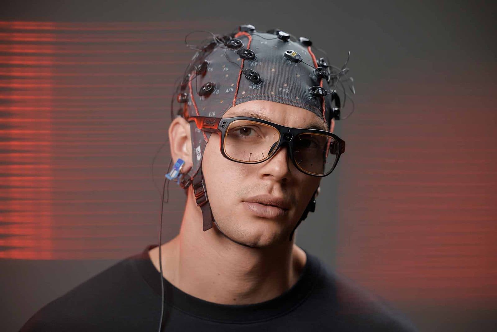
Synchronisation is the backbone of a multi-sensor study. To compare the moment gaze lands on a product with a change in EEG or EDA, all streams must be on the same timeline. There are three common ways to achieve this: devices recording on a shared clock (with g.Nautilus + Tobii Pro Glasses 3, gaze and EEG are recorded on the same clock), trigger markers sent from the stimulus computer to the devices, and network streaming protocols such as Lab Streaming Layer (LSL). Because of the display’s refresh delay, the moment an image actually appears on screen can differ from the moment the command was given; a light sensor measures that moment directly. Testing the timing during installation prevents surprises during analysis.
Limits and ethics: there is no “buy button”
The most often repeated misconception about neuromarketing is that the brain has a “buy button” and that, with the right measurement, a consumer’s decision can be read. Research shows nothing of the kind. In their review of the field’s hopes and hype, Ariely and Berns (2010) state that neuroimaging is not expected to become cheaper than conventional marketing methods in the near future, but that evidence is growing that it may provide information about the consumer experience not obtainable in other ways, and that its most promising use may come while a product is still just an idea. The main limits are these:
- The reverse inference problem: a change in a brain region or an EEG measure does not prove a specific mental process; the same activity can arise in many different processes (Poldrack 2006).
- Arousal is not valence: a rise in EDA or heart rate can indicate interest as well as discomfort.
- The gap between the lab and the real decision: how far a reaction measured in an experiment carries through to real purchases must be tested separately. Although EEG-based prediction studies report promising results, outcomes vary with method and context (Hakim and Levy 2019).
- Unpublished commercial claims: “neuro” metrics whose methods and data are not shared in peer-reviewed publications do not carry the same weight as verifiable scientific findings.
The ethical dimension matters as much as the measurement. Participants are informed about the purpose of the study and the data recorded, and they give consent; academic studies are conducted with ethics committee approval. EEG, eye tracking and physiological recordings are personal data and may fall within the special categories of personal data, such as health or biometric data, listed in Article 6 of Türkiye’s Law No. 6698 on the Protection of Personal Data (KVKK); storage, anonymisation and sharing should be planned with the institution’s data protection office. Studies with children and vulnerable groups require extra care. The aim is not to steer consumers but to understand how people actually perceive products and communication.
How to set up a neuromarketing lab
A neuromarketing or UX lab is not a single device but an arrangement that runs on one clock. Its typical components are these:
- Stimulus station
- A computer and display with known timing on which advertisements, web pages or product images are shown; in real-world studies, a shelf, a shop or a vehicle.
- Eye tracking
- A screen-based tracker for screen studies, and eye-tracking glasses with a scene camera for shelf and shop studies.
- EEG
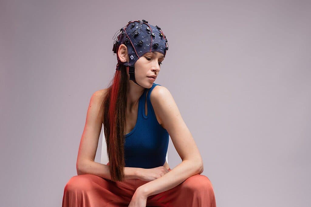
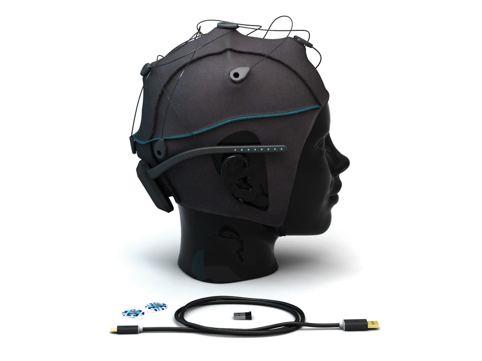
- For measures of attention and cognitive load; in the field, wireless systems that are quick to set up.
- Body signals
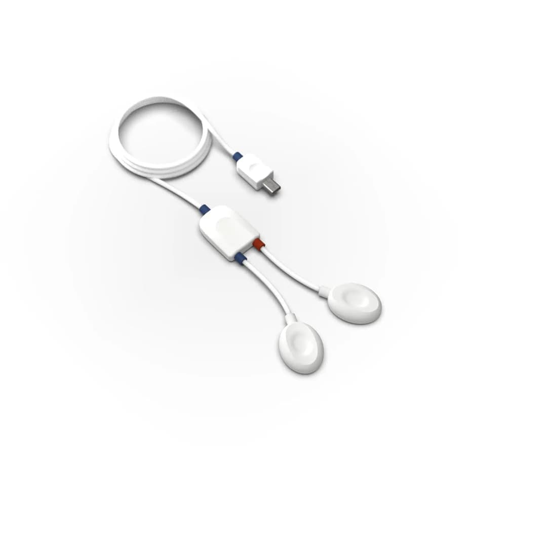
- EDA and heart rate (ECG or BVP), and respiration if needed; with a data acquisition hub at the screen and with wearable devices in the field.
- Synchronisation
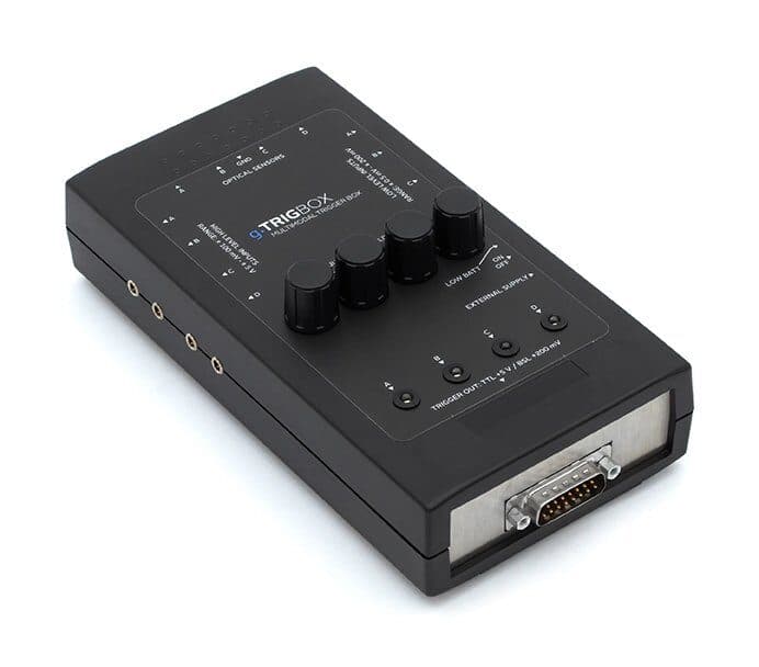
- A trigger box, a light sensor that captures the moment an image appears on screen, and a streaming infrastructure such as LSL.
- Setting
- A quiet room with steady temperature and lighting, and a separate workspace for the experimenter that does not disturb the participant.
- Analysis
- Recording and analysis software that brings the streams onto one timeline, and a team that knows the methods.
Nevosa’s UX and neuromarketing lab page describes configurations built from these components: a biosignal station that starts with skin conductance and heart rate; a lab that records research-grade EEG on the same clock as the biosignals; a real-world set-up that records in a shop or a vehicle with g.Nautilus EEG and Tobii Pro Glasses 3; and a neuroergonomics lab for driving and flight simulation. A lab can start small and grow on the same synchronisation infrastructure. Nevosa supplies these systems and provides installation, training, technical support and consulting from experimental design to data analysis. For planning, write your research question and the methods you want to use in the quote form.
Explore the animation
Follow, fixation by fixation, the gaze of a participant looking at a shop shelf: the scene and eye cameras of the eye-tracking glasses, the response of the visual cortex at the back of the head to each fixation, and the two streams joining on one timeline. It is a schematic drawing of how gaze is matched with the brain’s response in neuromarketing studies.
Where the eyes go, and how the brain answers
The glasses record what the participant looks at, the EEG how the brain responds to it. On one time axis, every look becomes a time mark in the brain signal. Switch the view, and tap the points in the drawing to see what each part does.
Infrared light makes small reflections on the cornea; eye cameras follow the pupil and these reflections, and where the gaze falls in the scene is calculated.
All notes
Fixation
A moment when the eye rests on one point. Visual information is taken in mostly during fixations; the larger the circle, the longer the gaze stayed there. The start of every fixation is a natural time mark for event-related analysis of the EEG.
- Typically 200–300 ms
- Taking in visual information
- A time mark for EEG
Saccade
The quick jump of the eye between two fixations. It usually lasts a few tens of milliseconds, and vision is largely suppressed meanwhile. At its start, a brief potential from the eye muscles can show in the EEG as a sharp deflection.
- A few tens of milliseconds
- Vision suppressed
- A sharp deflection in EEG
Scene camera
The camera at the front of the glasses records what the participant sees, from their point of view. The calculated gaze point is drawn onto this video, so you see exactly what the person looks at on a shelf, a screen or the road.
- Video from the participant's view
- Gaze point drawn onto the video
Eye cameras and infrared illuminators
Infrared illuminators in the frame make small reflections on the cornea; eye cameras follow the pupil and these reflections. Tobii Pro Glasses 3 builds a 3D eye model from corneal reflection, dark pupil and stereo geometry, and compensates slippage and parallax automatically.
- Corneal reflection · dark pupil
- 50 or 100 Hz, binocular
- 0.6° accuracy
The eye's electric field
The eyeball works like a small battery: the cornea is positive relative to the retina. As the eye turns, this field changes direction, and a blink does much the same. That makes eye movements one of EEG's strongest sources of artefacts, above all at electrodes near the eyes.
- Cornea positive, retina negative
- Eye movement and blink artefacts
Frontal electrode (Fp1)
The electrodes nearest the eyes see eye movements and blinks most strongly: the signal shifts as the eyes turn, and a blink makes a large, slow deflection. Because the eye-tracking data give the exact time of every movement and blink, these artefacts can be marked and removed from the EEG more reliably.
- Nearest the eyes
- Blink: a large, slow deflection
- Eye data mark the artefacts
The visual pathway
Light becomes a signal in the retina; the signal travels along the optic nerve, is relayed in the lateral geniculate nucleus of the thalamus (LGN), and reaches the primary visual cortex (V1) at the back of the head through the optic radiation. The journey takes a few tens of milliseconds, which is why the visual cortex's response to a fixation shows at occipital electrodes shortly after the fixation begins.
- Retina → optic nerve → LGN → V1
- A few tens of milliseconds
- Occipital lobe
Visual cortex (O1)
The visual cortex at the back of the head processes the visual information each new fixation brings. At occipital electrodes a positive wave appears about 80–100 ms after the fixation starts: the lambda response.
- Occipital region
- Lambda response
- About 80–100 ms after fixation
Synchronisation
EEG is recorded at 500 Hz, gaze at 50 or 100 Hz. g.HIsys Professional puts both streams on one time axis with millisecond precision, so it is known which EEG sample every fixation, saccade and blink falls on.
- EEG: 500 Hz
- Gaze: 50 / 100 Hz
- Millisecond synchronisation
Each dot is one gaze sample: 100 Hz, one every 10 ms. EEG is recorded five times as often, 500 samples a second; both sit on one time axis.
Frequently asked questions
What is neuromarketing?
Neuromarketing is the research field that studies how consumers respond to products, advertisements and brands through brain and body measurements such as EEG, eye tracking, electrodermal activity and fNIRS. In academia it is also called consumer neuroscience. Its aim is to measure reactions such as attention and arousal that self-report cannot capture.
Which devices are used in neuromarketing?
The most common methods are eye tracking, EEG and electrodermal activity; heart rate, fNIRS and facial expression analysis are also used. fMRI appears mostly in academic studies. For the results to be interpreted together, the devices must record on the same timeline.
What do you need to set up a neuromarketing lab?
You need a stimulus station with known timing, an eye-tracking system, sensors for body signals such as EEG, EDA and heart rate, and the trigger and streaming infrastructure that brings them onto one timeline. A quiet room with steady lighting and temperature, and a team that knows the methods, are also part of the lab. You can start small with a biosignal station and grow by adding EEG and eye tracking.
Can neuromarketing read a consumer’s mind?
No. The brain has no “buy button”; the measurements show changes in processes such as attention, arousal and cognitive load, and those changes can have many different causes. Findings are interpreted together with behavioural measures, at group level and in light of the experimental conditions.
What ethical rules apply to neuromarketing research?
Participants are informed about the purpose of the study and the data recorded, and they give consent; academic studies are conducted with ethics committee approval. Physiological recordings are personal data and may count as special-category personal data under Türkiye’s KVKK, so storage and sharing are planned accordingly. Studies with children and vulnerable groups require extra care.
References and further reading
- Plassmann et al. (2015) · Consumer Neuroscience: Applications, Challenges, and Possible Solutions
- Ariely and Berns (2010) · Neuromarketing: the hope and hype of neuroimaging in business
- Hakim and Levy (2019) · A gateway to consumers’ minds: EEG-based prediction in neuromarketing
- Dmochowski et al. (2014) · Audience preferences are predicted by temporal reliability of neural processing
- Poldrack (2006) · Can cognitive processes be inferred from neuroimaging data?
- Law No. 6698 on the Protection of Personal Data (KVKK, mevzuat.gov.tr, Turkish)