Neurotechnology glossary: English–Turkish terms
Neurotechnology glossary: English–Turkish terms for EEG, ERP, BCI, fNIRS, biosignals, TMS, tDCS, signal processing and synchronisation, with definitions.
Published
This neurotechnology glossary is a reference list that brings together the English and Turkish names, the abbreviations and short definitions of the core concepts in EEG, event-related potentials, brain–computer interfaces, fNIRS, biosignals, brain stimulation, signal processing and synchronisation. It is meant to help you quickly find the Turkish equivalent of a term, or what an English abbreviation stands for, while reading a paper, writing a thesis, or preparing a lab handout or a technical specification.
The terms are set out in eight groups, each written as “English term (Turkish term, abbreviation)”. Abbreviations keep the English form established in the literature: EEG, ERP, fNIRS, TMS. The Turkish equivalents are the ones used in the guides of this knowledge bank; where two Turkish terms are common in the literature, both are given. The definitions are written from a research-methods point of view; figures are quoted as the relevant guidelines or textbooks give them, as typical values. The glossary gives no diagnostic or treatment information; clinical uses are mentioned only as context, and any decision about an individual calls for a physician’s assessment. For more detail on a topic, see the knowledge bank’s guides to EEG, fNIRS, TMS, tDCS or the 10-20 system.
EEG and event-related potential (ERP) terms
Core concepts about the signal EEG measures, brain rhythms, event-related responses and electrodes:
- Electroencephalography (elektroensefalografi, EEG)
- A non-invasive method that follows the brain’s electrical activity on a millisecond scale by recording voltage differences between electrodes placed on the scalp. Much of the signal comes from the synchronous postsynaptic activity of populations of pyramidal neurons in the cortex.
- Frequency bands (frekans bantları; delta, theta, alpha, beta, gamma)
- The naming of rhythmic EEG activity by frequency. The IFCN glossary gives below 4 Hz for delta, 4–<8 Hz for theta, 8–13 Hz for alpha, 14–30 Hz for beta and above 30 Hz for gamma; studies report the limits they use.
- Alpha rhythm (alfa ritmi)
- An 8–13 Hz rhythm seen over the posterior regions of the head during wakefulness. It is most prominent with the eyes closed and the mind relatively idle, and is suppressed by eye opening, attention and mental effort.
- Mu rhythm (mu ritmi)
- A 7–11 Hz rhythm seen over the central regions. It decreases with movement of the opposite hand, with thinking of a movement or with readiness to move; motor imagery studies measure this decrease.
- Event-related potential (olaya ilişkin potansiyel, ERP)
- A change in potential obtained by averaging EEG segments time-locked to a stimulus, a response or a cognitive event. Averaging weakens the background activity that is not tied to the event; components are described by their latency and amplitude.
- Evoked potential (uyarılmış potansiyel, EP)
- The stimulus-locked response that a visual, auditory or somatosensory stimulus produces in the sensory pathways and cortex; referred to by the abbreviations VEP, AEP and SEP. ERP is the broader term and also covers components linked to cognitive processes such as attention or decision.
- P300 (P3)
- A positive ERP component, most prominent over parietal and central regions, that appears when a rare and task-relevant stimulus is noticed; it takes its name from its latency of about 300 ms. It is usually measured with the oddball paradigm, in which rare targets are presented among frequent standard stimuli.
- 10–20 system (10-20 sistemi)
- The international placement system in which electrode positions are defined at intervals of 10 and 20 per cent of the distances between the nasion, the inion and the two preauricular points. The letter gives the region (Fp, F, C, T, P, O), odd numbers the left and even numbers the right hemisphere, and “z” the midline; in the ACNS standard nomenclature the classic T3, T4, T5 and T6 positions are named T7, T8, P7 and P8 respectively.
- Reference electrode (referans elektrot)
- Every EEG channel is the potential difference between an electrode and a reference point; the reference is the common point of comparison for that difference. The mastoid or earlobe, Cz and an average reference computed in analysis are common choices; the choice changes the potential distribution and is therefore reported.
- Ground electrode (toprak elektrodu)
- The electrode that connects the participant to the amplifier’s common potential and helps suppress common-mode noise. It is not the same as the reference; channel values are not computed against it.
- Impedance (empedans)
- The opposition that the contact between electrode and skin presents to alternating current, measured in kiloohms (kΩ). High impedance, or impedance that differs between electrodes, increases noise; the target value is given in the manufacturer’s manual for the type of electrode and amplifier.
- Active electrode (aktif elektrot)
- An electrode that amplifies the signal at the point closest to its source, with a preamplifier inside the electrode, reducing the effect of cable movement and ambient interference. Electrodes without an amplifier are called passive electrodes.
- Dry and wet (gel) electrode (kuru ve ıslak ya da jelli elektrot)
- In a wet electrode, conductive gel provides a low and stable contact between electrode and skin. A dry electrode has no gel and a pin or conductive-polymer surface; it is quick to prepare and, because its contact impedance is higher, is usually designed as an active electrode.
- Artefact (artefakt)
- A signal of non-brain origin mixed into the recording: blinks and eye movements, muscle activity, heartbeat, sweating, electrode or cable movement and mains noise. Artefacts are divided into physiological and technical ones; removing them is a separate step of the analysis.
Brain–computer interface (BCI) terms
BCI paradigms, the parts of the loop and the measures that describe how well a system performs:
- Brain–computer interface (beyin bilgisayar arayüzü, BCI)
- A system that decodes signals measured from brain activity in real time and passes them as commands to a computer or device, without the need for muscle movement. It does not read thoughts; it recognises measurable responses produced by a given task or stimulus. The literature also uses the name brain–machine interface (beyin–makine arayüzü, BMI).
- Steady-state visually evoked potential (kararlı durum görsel uyarılmış potansiyel, SSVEP)
- The EEG response that appears over the occipital region at the frequency of a visual stimulus flickering at a constant rate, and at its harmonics, while the stimulus is looked at. Turkish sources also use the term “durağan durum”; in BCI it is used to determine which of several targets flickering at different frequencies is being looked at.
- Event-related desynchronisation and synchronisation (olaya ilişkin desenkronizasyon ve senkronizasyon, ERD/ERS)
- A decrease (ERD) or increase (ERS) in power in a given frequency band with an event, relative to a reference period before the event; expressed as a percentage change from that period. ERD in the mu and beta bands during movement or imagined movement, and ERS in the beta band after the movement ends, are typical examples.
- Motor imagery (motor imgeleme, MI)
- Imagining a movement without performing it. It produces mu and beta ERD over the sensorimotor cortex of the opposite hemisphere; it is used for self-paced BCI control that needs no external stimulus, and most users need training.
- P300 speller (P300 heceleyici)
- A BCI layout in which letters are shown in a matrix and rows and columns flash in random order. The P300 that appears when the row or column containing the target letter flashes is accumulated over many repetitions to determine the chosen letter (Farwell and Donchin, 1988).
- Synchronous and asynchronous BCI (senkron ve asenkron BCI; asynchronous also called self-paced)
- In a synchronous BCI the user gives commands only in time windows set by the system. An asynchronous, or self-paced, BCI must also work out by itself when the user wants to give a command; preventing false commands while the user rests is harder.
- Information transfer rate (bilgi aktarım hızı, ITR)
- The communication rate in bits per minute, calculated from accuracy, the number of classes and the selection time. When comparing BCIs it is reported together with accuracy, chance level and latency.
- Closed loop (kapalı döngü)
- An arrangement in which the decision drawn from the measured signal returns in real time to the participant or to a device. The participant sees the feedback and adjusts their strategy; offline accuracy does not stand in for control performance in the closed loop.
- Neurofeedback (nörofeedback)
- A closed-loop method in which the participant learns to regulate a feature of a brain signal, for example the power in an EEG band, with real-time feedback. In research it is used to study learning and self-regulation.
- Hybrid and passive BCI (hibrit ve pasif BCI)
- A hybrid BCI combines EEG with another signal, such as eye tracking, EMG or fNIRS, or combines two paradigms. A passive BCI monitors states such as workload or attention instead of taking commands.
fNIRS and optical brain imaging terms
Concepts relating to the hardware, physics and signal of measurement with near-infrared light:
- Functional near-infrared spectroscopy (fonksiyonel yakın kızılötesi spektroskopi, fNIRS)
- A non-invasive optical brain imaging method that follows changes in the concentration of oxygenated and deoxygenated haemoglobin in the cortex by measuring the part of near-infrared light, sent in through the scalp, that passes through the tissue and reaches the detectors. In Turkish tenders and specifications it also appears as “optik beyin görüntüleme sistemi” (optical brain imaging system).
- Optode (optot or optod)
- The common name for a light source or detector placed on the head. The arrangement of the optodes on a cap is called the optode geometry; publication recommendations ask for it to be reported in full.
- Source, detector and channel (kaynak, dedektör ve kanal)
- The source is an LED or laser diode emitting light at given wavelengths; the detector, for example a photodiode, measures the intensity of the light returning from the tissue. The measurement pair between one source and one detector is called a channel; the light follows a banana-shaped path between the two, and the region the channel measures lies roughly below the midpoint.
- Source–detector separation (kaynak–dedektör mesafesi)
- The distance between source and detector. As it increases the measurement becomes sensitive to greater depth, but less light reaches the detector; for standard channels in adults the typical value is about 3 cm, and shorter distances are used in infants.
- Short-separation channel (kısa mesafe kanalı)
- A channel formed by a detector very close to the source, which measures almost only the changes in the scalp and skull. Yücel et al. (2021) count channels shorter than 15 mm in this class and recommend about 8 mm for adults; its signal is used to remove the superficial component from the long channels.
- Oxygenated and deoxygenated haemoglobin (oksijenli ve oksijensiz hemoglobin, HbO/HbR)
- The two chromophores whose concentration changes fNIRS measures; the literature also uses HbO2 and HHb, or oxyhaemoglobin and deoxyhaemoglobin. Their absorption curves cross at about 800 nm (the isosbestic point), so systems use at least two wavelengths, one below and one above that point.
- Haemodynamic response (hemodinamik yanıt)
- The change in blood flow and oxygenation that accompanies neural activity; the link between neural activity and local blood flow is called neurovascular coupling (nörovasküler eşleşme). After a brief stimulus the response typically begins within 1–2 s, peaks at about 5–6 s and returns to baseline within 15–20 s; the expected pattern is a rise in HbO accompanied by a smaller fall in HbR.
- Modified Beer–Lambert law (değiştirilmiş Beer–Lambert yasası, MBLL)
- The relation that links the change in optical density measured at each wavelength to the concentration changes of HbO and HbR; it includes the absorption coefficients, the source–detector separation and the differential pathlength factor. In continuous-wave systems the result is a relative change, not an absolute one.
- Differential pathlength factor (diferansiyel yol uzunluğu faktörü, DPF)
- The ratio of the mean path that light travels in tissue, lengthened by scattering, to the source–detector separation. It is taken from the literature by age and wavelength, and the value used is reported.
- Continuous-wave, frequency-domain and time-domain fNIRS (sürekli dalga, frekans alanı ve zaman alanı fNIRS; CW, FD, TD)
- Continuous-wave systems measure only light intensity and give relative changes; most commercial systems are of this type. Frequency-domain and time-domain systems also measure the phase shift or time of flight of the light and can estimate the optical properties of the tissue.
- High-density diffuse optical tomography (yüksek yoğunluklu difüz optik tomografi, HD-DOT)
- A method that combines overlapping measurements at several distances, taken from many sources and detectors set in a dense grid, with a model of light propagation to produce a three-dimensional map of haemoglobin changes.
Biosignal terms: EMG, ECG, EDA, PPG, respiration and the eyes
A biosignal is a physiological signal measured from a living organism that changes over time; it may be electrical (EMG, ECG, EDA), optical (PPG) or mechanical (respiration, movement) in origin. The terms in this group concern measurements of muscle, heart, skin, breathing and the eyes:
- Surface electromyography (yüzey elektromiyografisi, sEMG)
- The recording of the electrical activity of muscle fibres with electrodes on the skin, as distinct from needle EMG, which uses electrodes inserted into the muscle. In research the SENIAM recommendations are widely used for electrode placement and skin preparation.
- Motor unit action potential (motor ünite aksiyon potansiyeli, MUAP)
- A motor neuron and all the muscle fibres it innervates form a motor unit; when the motor neuron fires, the electrical wave that travels along the fibres is the MUAP. Surface EMG measures not a single unit but the sum of many MUAPs under the electrode.
- Maximum voluntary contraction (maksimal istemli kasılma, MVC)
- The largest voluntary contraction a participant can produce in a muscle under standard conditions. EMG amplitudes are usually normalised as a percentage of MVC (%MVC) so that they can be compared between people and sessions; amplitude itself is usually summarised with a linear envelope or RMS.
- Electrocardiography (elektrokardiyografi, ECG)
- The recording of the heart’s electrical activity with electrodes on the skin; the common Turkish abbreviation is EKG. In research the R peaks are usually detected, and heart rate and the RR intervals between successive beats are derived.
- Heart rate variability (kalp hızı değişkenliği, HRV)
- The variation over time of the interval between successive heartbeats. It is summarised in the time domain with measures such as SDNN and RMSSD, and in the frequency domain with the power in the low-frequency (LF, 0.04–0.15 Hz) and high-frequency (HF, 0.15–0.4 Hz) bands.
- Electrodermal activity (elektrodermal aktivite, EDA)
- The change in the electrical properties of the skin that follows the activity of sweat glands controlled by the sympathetic nervous system; usually measured as skin conductance, in microsiemens (µS). Its older and still common name is galvanic skin response (galvanik deri tepkisi, GSR).
- Skin conductance level and response (deri iletkenlik düzeyi ve yanıtı, SCL/SCR)
- SCL is the tonic baseline of EDA, changing slowly over minutes. An SCR is a phasic rise that typically begins 1–3 s after a stimulus, peaks within a few seconds and decays more slowly; those that occur without a visible stimulus are called spontaneous or non-specific SCRs (NS-SCR).
- Photoplethysmography (fotopletismografi, PPG)
- An optical method that measures changes in blood volume in the microcirculation from the change in absorption of light sent into the tissue; the pulse wave and pulse rate are derived from this signal. Some sensors carry it in their name as blood volume pulse (kan hacmi nabzı, BVP).
- Respiration recording (solunum kaydı; RIP, PZT)
- Methods that measure the change in volume with breathing using a belt around the chest or abdomen: respiratory inductance plethysmography (endüktif pletismografi, RIP) and piezoelectric (PZT) sensors. Respiration is studied in its own right and also recorded to track the effect of breathing in EDA, HRV and fNIRS data.
- Electrooculography (elektrookülografi, EOG)
- A method that measures eye movements and blinks with electrodes around the eyes, using the potential difference between the cornea and the retina. It is also recorded alongside EEG to track and remove eye artefacts.
- Eye tracking (göz takibi)
- A method that measures where and when the gaze is directed; video-based systems usually follow the pupil and the corneal reflection under infrared illumination. There are screen-based and wearable (glasses) systems; Turkish also uses the term “göz izleme”.
- Fixation (fiksasyon)
- A period in which the gaze stays relatively still at one point for a short time; visual information is taken in largely during fixations. A fixation typically lasts 200–300 ms.
- Saccade (sakkad)
- A rapid eye movement that carries the gaze from one point to another; it usually lasts a few tens of milliseconds, and visual perception is largely suppressed during it. Studying the speed and duration of saccades requires a high sampling rate.
- Area of interest (ilgi alanı, AOI)
- A region of the stimulus defined for analysis, for example a logo or a product. Measures such as time to first fixation, dwell time and number of fixations are computed per AOI.
Brain stimulation terms: TMS, tDCS and FES
Concepts relating to magnetic and electrical stimulation methods, their protocols and control conditions. These terms describe research protocols; clinical applications depend on a physician’s assessment.
- Transcranial magnetic stimulation (transkraniyal manyetik stimülasyon, TMS)
- A non-invasive stimulation method that induces an electric field in brain tissue through the changing magnetic field created by a brief current pulse in a coil held over the head. At sufficient intensity it can fire neurons directly; figure-of-eight (butterfly) coils produce a focal field, circular coils a broader one.
- Motor evoked potential (motor uyarılmış potansiyel, MEP)
- The response produced in the target muscle on the opposite side by stimulation of the motor cortex and recorded with surface EMG. Its peak-to-peak amplitude is a measure of corticospinal excitability and its latency of conduction; because amplitude varies markedly from trial to trial, many trials are averaged.
- Motor threshold (motor eşik; RMT, AMT)
- The resting motor threshold (RMT) is, by the conventional definition, the lowest stimulator output that produces an MEP above about 50 µV in at least 5 of 10 consecutive stimuli with the muscle relaxed. The active motor threshold (AMT) is determined during a slight contraction and is lower; the intensity of other protocols is given as a percentage of this threshold.
- Repetitive TMS (tekrarlayıcı TMS, rTMS)
- A protocol in which pulses are delivered in trains at a set frequency. In group averages, low-frequency (about 1 Hz) rTMS tends to decrease cortical excitability and high-frequency (5 Hz and above) rTMS to increase it; the effects vary markedly between individuals.
- Theta-burst stimulation (theta-burst stimülasyonu, TBS; cTBS, iTBS)
- A form of rTMS in which bursts of three pulses at 50 Hz are repeated five times a second. In the motor cortex, continuous TBS (cTBS) tends to decrease excitability and intermittent TBS (iTBS) to increase it (Huang et al., 2005).
- Neuronavigation (nöronavigasyon)
- A targeting system that tracks the position and orientation of the coil in real time on the participant’s MRI or on a template brain. It makes it possible to return to the same target between sessions and to report the actual stimulation sites.
- TMS–EEG and TMS-evoked potential (TMS–EEG ve TMS ile uyarılmış potansiyel, TEP)
- The simultaneous EEG recording of the cortical response to TMS pulses; the pulse-locked response is called a TEP. It requires TMS-compatible amplifiers and electrodes; pulse, muscle, auditory and somatosensory components are separated from the cortical response.
- Transcranial direct current stimulation (transkraniyal doğru akım uyarımı, tDCS)
- A method that temporarily changes cortical excitability by passing a weak, constant direct current between electrodes on the scalp. It does not fire neurons directly but shifts the membrane potential and so changes the likelihood of firing; current intensity (mA), electrode size, current density (mA/cm²), duration and montage are reported.
- Anodal and cathodal stimulation (anodal ve katodal uyarım)
- The electrode where the current enters is the anode, the one where it leaves the cathode. In the foundational studies over the motor cortex, anodal stimulation tends to increase excitability and cathodal stimulation to decrease it; the effect can vary with intensity, duration and the individual.
- Transcranial alternating current stimulation (transkraniyal alternatif akım uyarımı, tACS)
- Stimulation with a current oscillating at a set frequency, intended to interact with brain rhythms. Together with tDCS and transcranial random noise stimulation (tRNS) it belongs to the family of transcranial electrical stimulation (tES).
- Sham stimulation (sham, yalancı uyarım)
- A control condition that imitates the sensation of real stimulation. In tDCS the current is ramped up, held at the target intensity only briefly and ramped down; in TMS a sham coil is used. The aim is to separate the effect from expectation and placebo, and the success of blinding is checked at the end of the session.
- Functional electrical stimulation (fonksiyonel elektriksel stimülasyon, FES)
- A method that produces a functional muscle contraction by stimulating peripheral nerves or muscles with electric current. In BCI research it is used in closed-loop arrangements triggered by the detection of motor imagery.
Signal processing and analysis terms
The steps of the pipeline from raw recording to result, and classification methods:
- High-pass, low-pass and band-pass filter (yüksek, alçak ve bant geçiren filtre)
- Operations that attenuate frequencies below a cut-off, above it, or outside a range. The cut-off frequency, filter type and order are reported, because a filter can also change the shape of responses in the time domain.
- Notch filter (çentik filtre)
- A filter that suppresses a narrow frequency range, usually mains noise; the mains frequency in Türkiye is 50 Hz. If used, it is reported; methods that estimate and subtract the mains component are another option.
- Re-referencing and average reference (yeniden referanslama ve ortalama referans)
- Converting the recording reference to another reference in analysis. With an average reference, the mean of all channels at each moment is subtracted from every channel.
- Independent component analysis (bağımsız bileşen analizi, ICA)
- A method that separates a multichannel signal into statistically independent components. It is widely used to isolate and remove artefacts such as blinks, eye movements and the heartbeat; which components were removed, and why, is reported.
- Epoch and epoching (epok ve epoklama)
- Cutting the continuous signal into segments according to the event markers, with a set window before and after each event; each segment is called an epoch. It is a basic step of ERP and time–frequency analysis.
- Baseline correction (bazal dönem düzeltmesi)
- Subtracting the mean of a pre-event reference window from each epoch; ERP amplitudes are measured relative to this period. The length of the window is reported.
- Power spectral density (güç spektral yoğunluğu, PSD)
- The distribution of signal power over frequency. In EEG it is usually estimated with Welch’s method, by averaging the Fourier transforms of overlapping windows; it is the basis of band power in resting-state recordings.
- Time–frequency analysis (zaman–frekans analizi)
- Analysis that shows how power changes over frequency and time together; Morlet wavelets or the short-time Fourier transform are commonly used. ERD/ERS maps are obtained with this analysis.
- Common spatial patterns (ortak uzamsal örüntüler, CSP)
- A method that finds spatial filters maximising the variance in one class while minimising it in the other. In motor imagery BCIs it is a common feature extraction step, for example to separate imagined left-hand and right-hand movement.
- Linear discriminant analysis (doğrusal ayırtaç analizi, LDA)
- A classifier that separates classes with a linear decision boundary; in Turkish it is also called “doğrusal diskriminant analizi”. Because it is simple and computationally light, it is widely used in real-time BCIs.
- Cross-validation (çapraz doğrulama)
- Estimating how well a model performs on data it has not seen by repeatedly splitting the data into training and test parts. Preprocessing and feature selection are fitted on the training data only; otherwise information leaks from the test data.
- Source localisation (kaynak konumlandırma)
- Estimating the location of cortical sources from scalp measurements. It requires a head model, electrode coordinates and a solution to the inverse problem, and carries uncertainty; the highest point of a topographic map is not the location of the source.
Hardware, synchronisation and data format terms
Terms about amplifier properties, event timing and data storage, which also appear in technical specifications:
- Sampling rate (örnekleme hızı)
- The number of samples taken per second (Hz). To avoid aliasing, the Nyquist principle requires it to be more than twice the highest frequency in the signal; in practice it is chosen much higher because of filtering and analysis needs.
- Analogue-to-digital resolution (analog-dijital çözünürlük, ADC resolution)
- The number of bits with which the analogue signal is digitised; research amplifiers usually use 24 bits. The smallest change that can actually be resolved is set by the input range and noise as well as by the number of bits.
- Input noise (giriş gürültüsü)
- The amplifier’s own noise, measured with its inputs shorted. It is given in µV, stating whether it is RMS or peak-to-peak and over which bandwidth it was measured.
- Common-mode rejection ratio (ortak mod reddi oranı, CMRR)
- A ratio that shows how strongly a differential amplifier suppresses noise common to both inputs, such as mains interference. It is given in decibels (dB), stating the frequency at which it was measured.
- DC coupling and input range (DC kuplaj ve giriş aralığı)
- DC-coupled inputs also record slow components down to zero hertz; AC-coupled inputs filter them out in hardware. The input range is the largest voltage the amplifier can measure without saturating; it matters for carrying large artefacts without saturation when recording together with tDCS or TMS.
- Trigger and event marker (tetik ve olay işareti)
- A trigger is a signal that writes the moment of a stimulus or response into the recording; it is usually sent as a TTL pulse from the stimulus computer or a trigger box to the amplifier’s digital input. The timestamp that identifies an event by its code in the recording is called an event marker; the number of bits of the trigger input determines how many distinct event codes can be told apart.
- Latency and jitter (gecikme ve zamanlama sapması)
- The constant difference between the trigger and the actual moment of the stimulus is latency; the difference that varies from trial to trial is jitter. Both are measured with a photodiode placed on the stimulus screen; constant latency can be corrected in analysis, whereas jitter smears and shrinks the averaged ERP.
- Lab Streaming Layer (LSL)
- An open-source software layer that aligns data streams and event markers from different devices to a common clock over the network. It is widely used to combine EEG, eye tracking and biosignals in the same session; the timing accuracy achieved is verified by measurement in the set-up.
- EDF and BDF (European Data Format, BioSemi Data Format)
- Common open file formats for biosignals. EDF is a 16-bit format and BDF its 24-bit adaptation; EDF+ can also store event markers.
- BIDS (Brain Imaging Data Structure)
- A community standard for the folder structure, file names and metadata of neuroimaging and electrophysiology data. There is an EEG-BIDS extension for EEG (Pernet et al., 2019); it makes sharing data and repeating analyses easier.
Research design and practice terms
Concepts of experimental design, the ethics process and reporting that are shared by every method:
- Paradigm (paradigma)
- The experimental design that defines what the participant will do, which stimuli are presented in which order and which signal feature is measured; oddball, SSVEP and motor imagery are each a paradigm.
- Trial and condition (deneme ve koşul)
- A trial is one presentation of a stimulus or task; a condition is each of the experimental states being compared. ERP and fNIRS analyses rest on the average of many trials of each condition.
- Block design and event-related design (blok tasarımı ve olay ilişkili tasarım)
- In a block design the trials of one condition are grouped in blocks lasting tens of seconds; in an event-related design trials are presented one at a time and in mixed order. Because the haemodynamic response is slow, event-related fNIRS designs keep long and variable intervals between trials.
- Control condition and blinding (kontrol koşulu ve körleme)
- A control condition resembles the experimental condition as closely as possible apart from the variable under study. With blinding, the participant, and in double-blind studies the assessor too, does not know which condition is being run.
- Ethics committee approval and informed consent (etik kurul onayı ve bilgilendirilmiş onam)
- The approval of the protocol of research with human participants by an independent ethics committee, and the participant’s written statement of voluntary participation after understanding its purpose, method and possible risks.
- Safety screening (güvenlik taraması)
- Screening participants with a standard questionnaire before stimulation studies. For TMS, a history of epilepsy, medications that may lower the seizure threshold, and intracranial metal or electronic implants are asked about; the decision rests on the ethics-approved protocol and, where needed, a physician’s assessment.
- Multimodal recording (çok modlu kayıt)
- Recording more than one measure, such as EEG, fNIRS, eye tracking and biosignals, in the same session on a common time base. It is interpreted bearing in mind that each method sees a different time scale and physiological process.
- Hyperscanning
- Recording two or more people simultaneously with EEG, fNIRS or another method during an interaction. It is used to study the coupling of brain activity between people and requires the recordings to share a common clock; Turkish publications mostly use the English name.
- Multiple comparisons correction (çoklu karşılaştırma düzeltmesi)
- Methods that control the false-positive rate of tests run over many channels, time points or frequencies: the Bonferroni correction, the false discovery rate (yanlış keşif oranı, FDR) and cluster-based permutation tests.
- Reproducibility (tekrar üretilebilirlik)
- The ability to reach the same result with the same data and methods. Complete reporting of filters, reference, excluded data, software versions and parameters, and sharing data in standards such as BIDS, support it.
Commonly confused pairs of terms
The pairs below are often confused in papers and specifications. The table sums up, in one row each, the difference between the two terms and why it matters:
| Pair of terms | Difference | Why it matters |
|---|---|---|
| Reference electrode / ground electrode | The reference is every channel’s point of comparison; the ground helps suppress common noise, and channel values are not computed against it. | The choice of reference changes the potential distribution and is reported. |
| Event-related potential (ERP) / evoked potential (EP) | An EP is the response of the sensory pathways to a sensory stimulus; ERP is the broader term that also covers components linked to cognitive processes. | The P300 is an ERP component; the visual evoked potential (VEP) is an EP. |
| ERD / ERS | ERD is a decrease in band power, ERS an increase; both are expressed as a percentage of a pre-event reference period. | Motor imagery BCIs classify mu and beta ERD. |
| SCL / SCR | SCL is the tonic level that changes over minutes; an SCR is a phasic rise beginning 1–3 s after a stimulus. | They answer different questions and are reported separately. |
| HbO / HbR | With activity HbO rises and HbR falls with a smaller amplitude. | Both changing in the same direction can signal a systemic effect or movement; they are reported together. |
| Resting motor threshold (RMT) / active motor threshold (AMT) | RMT is determined with the muscle relaxed, AMT during a slight contraction; AMT is lower. | Protocol intensity is written stating which threshold it is a percentage of. |
| Sampling rate / analogue-to-digital resolution | Sampling rate sets resolution in time, ADC resolution resolution in amplitude. | They are separate items in a specification; one does not replace the other. |
| Latency / jitter | Latency is a constant difference and can be corrected; jitter varies from trial to trial. | Jitter smears the averaged ERP; both are measured with a photodiode. |
| Accuracy / precision (eye tracking) | Accuracy is the mean deviation of the measured gaze from the true gaze point; precision is how close successive samples are to each other. | AOI size is chosen according to accuracy; low precision makes fixation detection harder. |
| Trigger / Lab Streaming Layer (LSL) | A trigger writes the moment of an event into the recording over a hardware line; LSL aligns data streams and markers to a common clock over the network. | In multi-device set-ups the two can be used together; timing is verified by measurement in every case. |
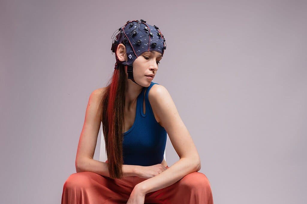
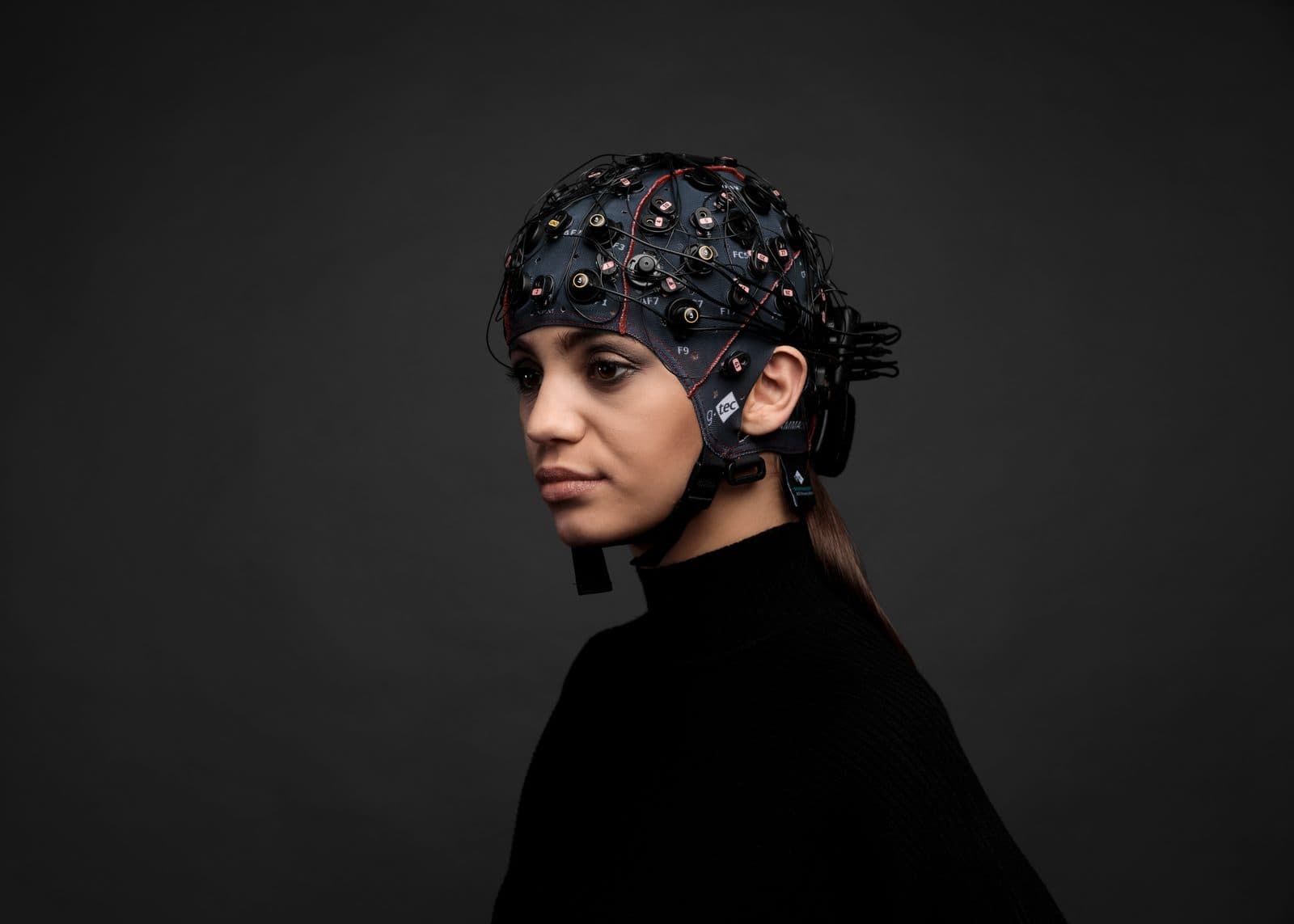
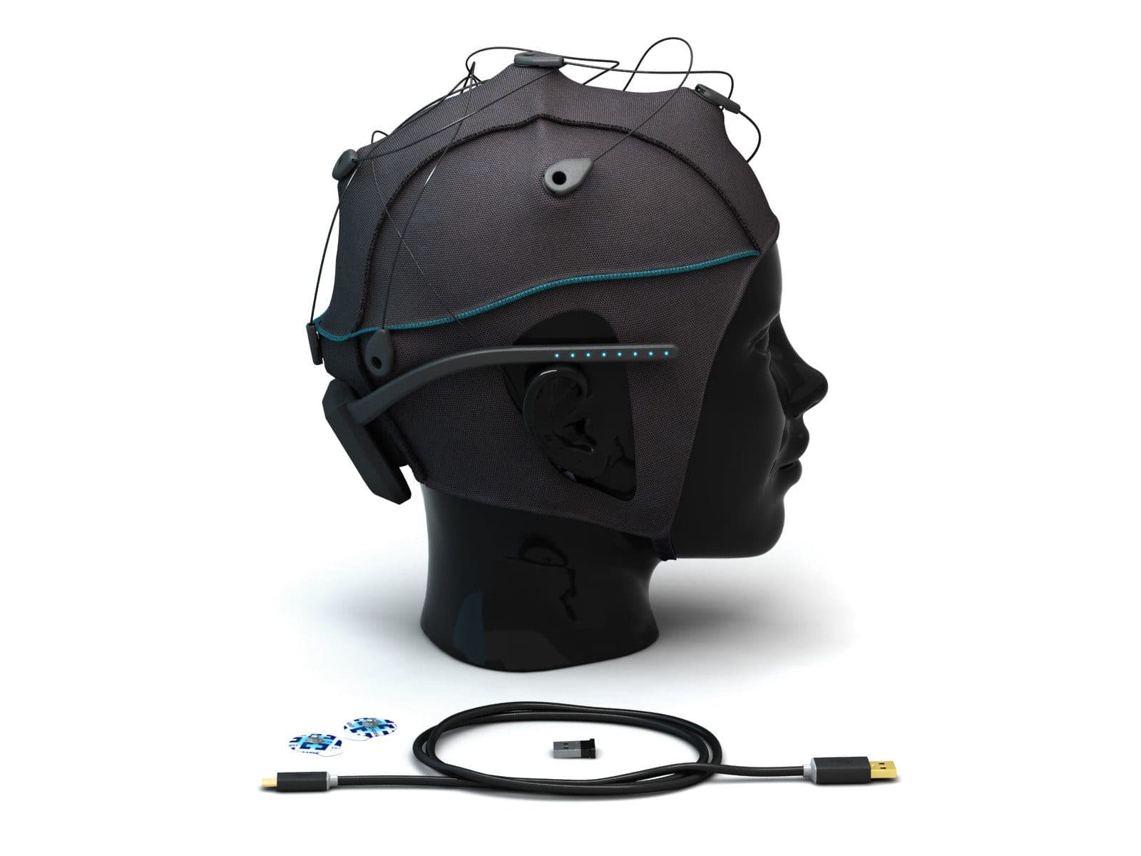
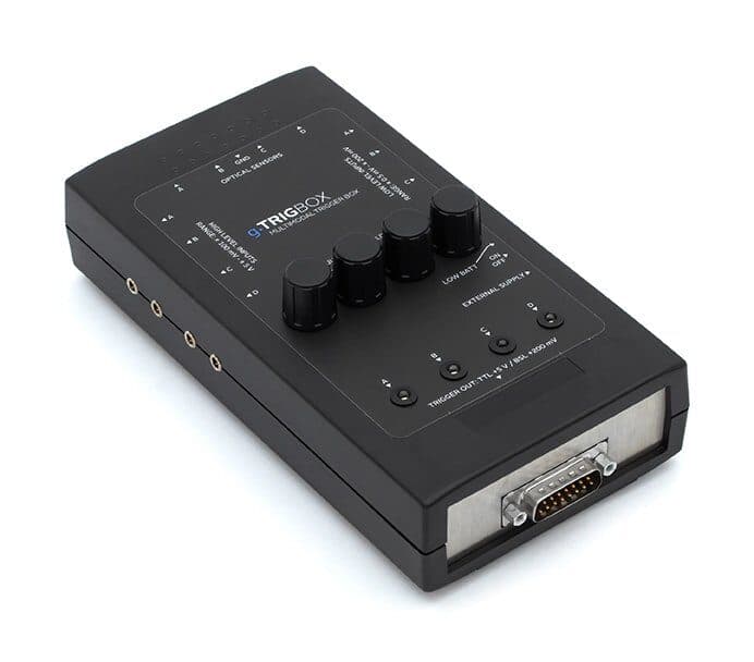
Some of the measurements named in this glossary are made with systems in Nevosa’s catalogue: g.Nautilus Research for EEG, g.Nautilus fNIRS for recording EEG and fNIRS in the same cap, Unicorn Hybrid Black for BCI studies, biosignalsplux for biosignal sensors and g.TRIGbox for generating triggers. The details are on the product pages; for installation, training and technical support you can use the quote form.
Frequently asked questions
What does SSVEP mean?
SSVEP stands for steady-state visually evoked potential; in Turkish it is called “kararlı durum” (or “durağan durum”) görsel uyarılmış potansiyel. It is the EEG response that appears over the occipital region at the frequency of a visual stimulus flickering at a constant rate, and at its harmonics, while the stimulus is looked at. In brain–computer interfaces it is used to determine which of several targets flickering at different frequencies is being looked at.
What is an optode?
An optode (optot or optod in Turkish) is the common name for a light source or detector placed on the head in an fNIRS measurement. The source emits near-infrared light and the detector measures the light returning from the tissue; the measurement pair between one source and one detector is called a channel. The arrangement of the optodes on the cap is reported as the optode geometry.
What are ERD and ERS?
ERD (event-related desynchronisation) is the decrease in power in a given frequency band with an event, relative to a reference period before the event; ERS (event-related synchronisation) is the corresponding increase. Both are expressed as a percentage change from the reference period. For example, ERD appears in the mu and beta bands during a hand movement or imagined movement, and ERS in the beta band after the movement ends.
What is the difference between an ERP and an evoked potential?
An evoked potential (EP) is the stimulus-locked response that a visual, auditory or somatosensory stimulus produces in the sensory pathways and cortex. An event-related potential (ERP) is the broader term; it also covers components linked to cognitive processes such as attention, expectation or decision. The P300, for example, is an ERP component, while the visual evoked potential (VEP) is an EP.
Are the reference and ground electrodes the same thing in EEG?
No. The reference electrode is the common point of comparison against which each channel’s potential difference is computed, and its choice changes how the data look. The ground electrode connects the participant to the amplifier’s common potential and helps suppress common-mode noise; channel values are not computed against it.
References and further reading
- Kane et al. (2017) · A revised glossary of terms most commonly used by clinical electroencephalographers, revision 2017 · Clinical Neurophysiology Practice
- Acharya et al. (2016) · ACNS Guideline 2: Guidelines for standard electrode position nomenclature · Journal of Clinical Neurophysiology
- Pfurtscheller and Lopes da Silva (1999) · Event-related EEG/MEG synchronization and desynchronization: basic principles · Clinical Neurophysiology
- Yücel et al. (2021) · Best practices for fNIRS publications · Neurophotonics
- Rossini et al. (2015) · Non-invasive electrical and magnetic stimulation of the brain, spinal cord, roots and peripheral nerves: an updated IFCN committee report · Clinical Neurophysiology
- Antal et al. (2017) · Low intensity transcranial electric stimulation: safety, ethical, legal regulatory and application guidelines · Clinical Neurophysiology