What is TMS? From magnetic stimulation to TMS–EEG research
Learn how transcranial magnetic stimulation works and compare clinical TMS, research TMS and TMS–EEG with an interactive animation.
What does TMS change?
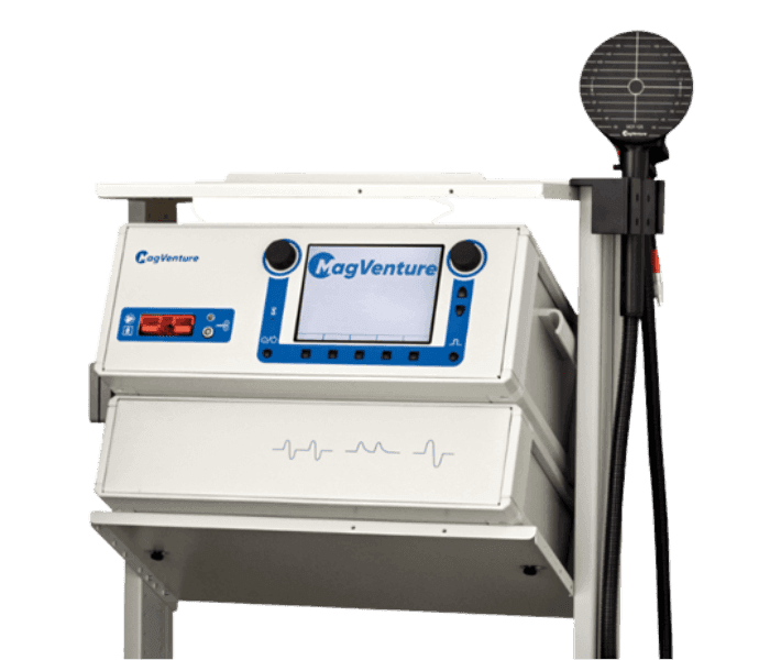
Transcranial magnetic stimulation (TMS) uses brief currents in a coil outside the head to create a changing magnetic field. The electric field induced in tissue can affect neural activity under appropriate conditions. TMS stimulates rather than records; EEG can record responses when the methods are combined. Coil geometry, position, orientation and individual anatomy influence the stimulation field.
Clinical and research TMS
Clinical TMS is delivered within specific indications and the device’s approved scope, following medical assessment. Research TMS investigates motor cortical excitability, causal brain–behaviour relationships and network dynamics. Single-pulse, paired-pulse and repetitive TMS (rTMS) are distinct experimental approaches with different purposes.
Planning a research session
The research question must relate clearly to the measured outcome. Motor studies may measure electromyographic motor evoked potentials (MEPs), whereas TMS–EEG studies assess EEG responses alongside their sensory contributions.
- Define the research question, target and control condition.
- Complete ethics approval, consent and participant safety screening.
- Choose the coil and recording equipment according to their instructions.
- Document targeting, coil orientation and reproducibility of positioning.
- Synchronise stimulation markers with EEG or EMG recordings.
- Report controls, artefacts and outcome measures.
Interpreting TMS–EEG
A large signal immediately after stimulation cannot automatically be attributed to a cortical response. Pulse artefacts, muscle activity, electrode movement and auditory or somatosensory responses may contribute. Artefact handling and suitable controls are essential when interpreting TMS-evoked potentials (TEPs). Selecting MagVenture Turkey or TMS–EEG research equipment should start with the technical requirements of the intended study.
Explore the animation
Explore the relationship between coil current, induced electric field and example recordings. Traces are illustrative and do not constitute a clinical protocol.
From a magnetic pulse to a brain response.
Transcranial magnetic stimulation (TMS) uses a coil positioned over the head to generate a rapidly changing magnetic field. This induces an electric field in cortical tissue that can affect neural activity. Single pulses, repeated pulse trains and simultaneous EEG address different questions.
Orientation, distance and tissue geometry affect field distribution. Arrows illustrate orientation schematically.
Coil current I(t)
Induced field E(t)
E(t) ~ -dI(t)/dt
For fixed coil geometry, the induced-field time course relates to the rate of change of coil current. The curves are separately normalized illustrations, not device outputs or individualized field calculations.
Stimulation → recording → analysis
Schematic illustration · Not a clinical dose or real patient recording.
A reproducible clinical workflow.
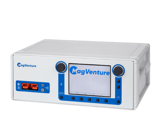
For clinical rTMS and theta burst (TBS), system selection depends on the clinician-defined protocol, coil, cooling and session workflow. Clinical use depends on the approved indications of the device and configuration.
- Motor threshold assessment
- Coil positioning
- Protocol and session tracking
Investigate cortical circuits through your experimental design.
For motor evoked potentials (MEP), cortical excitability, motor mapping and paired-pulse studies, consider pulse shape, current direction, triggering and coil options together. MagPro X100 and the MagOption configuration offer options for advanced research workflows.
- MEP and motor mapping
- Paired-pulse protocols
- External triggering and synchronization
Observe cortical responses following magnetic stimulation.
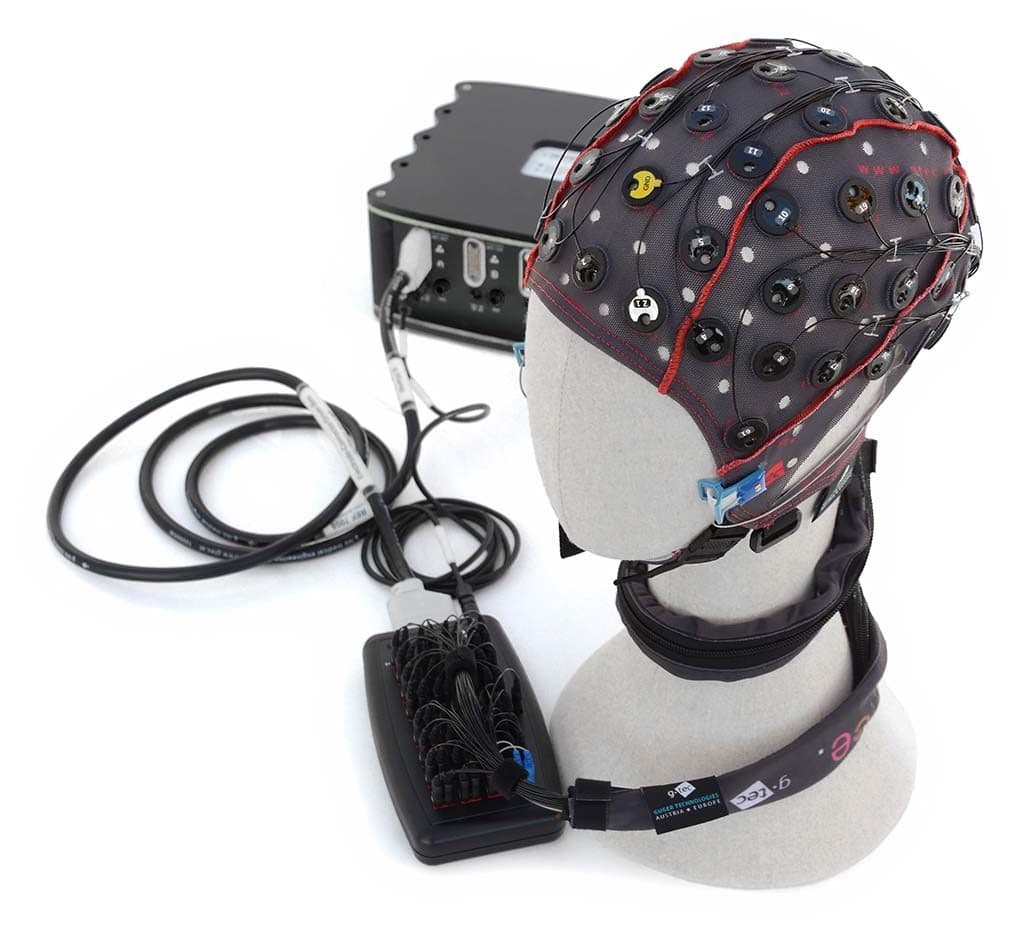
TMS–EEG combines stimulation with simultaneous EEG acquisition to study TMS-evoked potentials (TEP) and their temporal distribution. A setup with g.LADYbird TMS–EEG electrodes and g.HIamp must address triggering, pulse artifacts, muscle responses and auditory/somatosensory effects in its analysis plan.
- TMS-compatible EEG electrodes
- Shared event timing
- TEP and artifact analysis
- 01
Triggering and raw recording
Align EEG/EMG with TMS events; define the pre-stimulation reference and response windows.
- 02
Artifacts and confounding effects
Control pulse artifacts, muscle activity, electrode effects and auditory/sensory responses. Indiscriminate filtering can alter the response itself.
- 03
Quantitative response assessment
Compare MEP amplitude and latency for TMS–EMG, or TEP, time–frequency and topographic distributions for TMS–EEG across experimental conditions.
- 01
A current pulse in the coil
The stimulator sends a brief high-current pulse through the coil.
- 02
Magnetic field and induction
The changing magnetic field induces an electric field in tissue; its effect depends on coil geometry and orientation.
- 03
Evaluate the response
Depending on the study, assess motor responses with EMG, cortical responses with EEG or clinical outcomes with appropriate assessment tools.