Skip to content
g.tec medical engineering · g.Nautilus series

g.Nautilus fNIRS

Wearable EEG and fNIRS

A wearable system that combines simultaneous EEG and fNIRS recording in a single cap. EEG shows when brain activity happens, to the millisecond; fNIRS shows where, to the millimetre. Together they give a comprehensive analysis at high temporal and spatial resolution.

  • 8 / 16 / 32 / 64 EEG channels
  • 16 sources + 16 detectors
  • 760 and 850 nm
  • g.HIsys integration
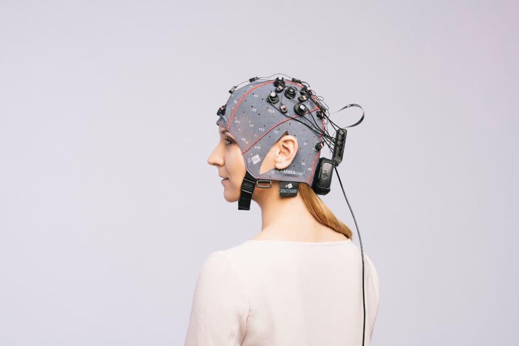
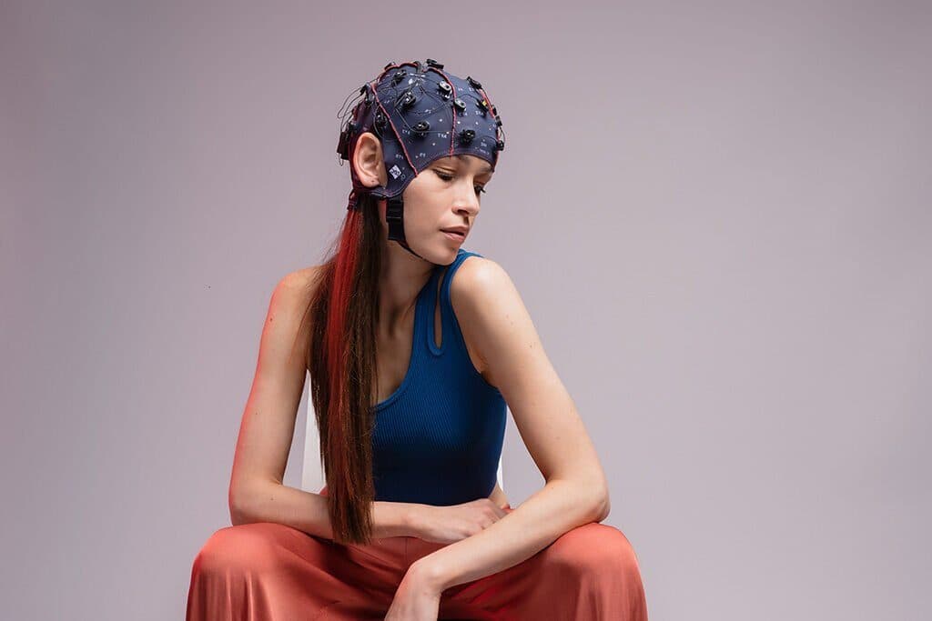
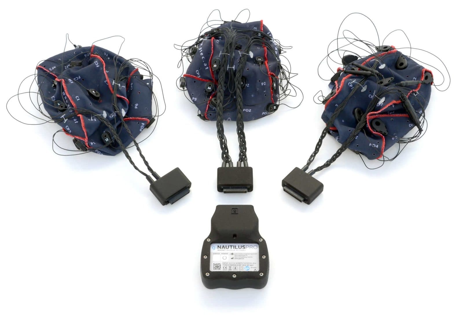
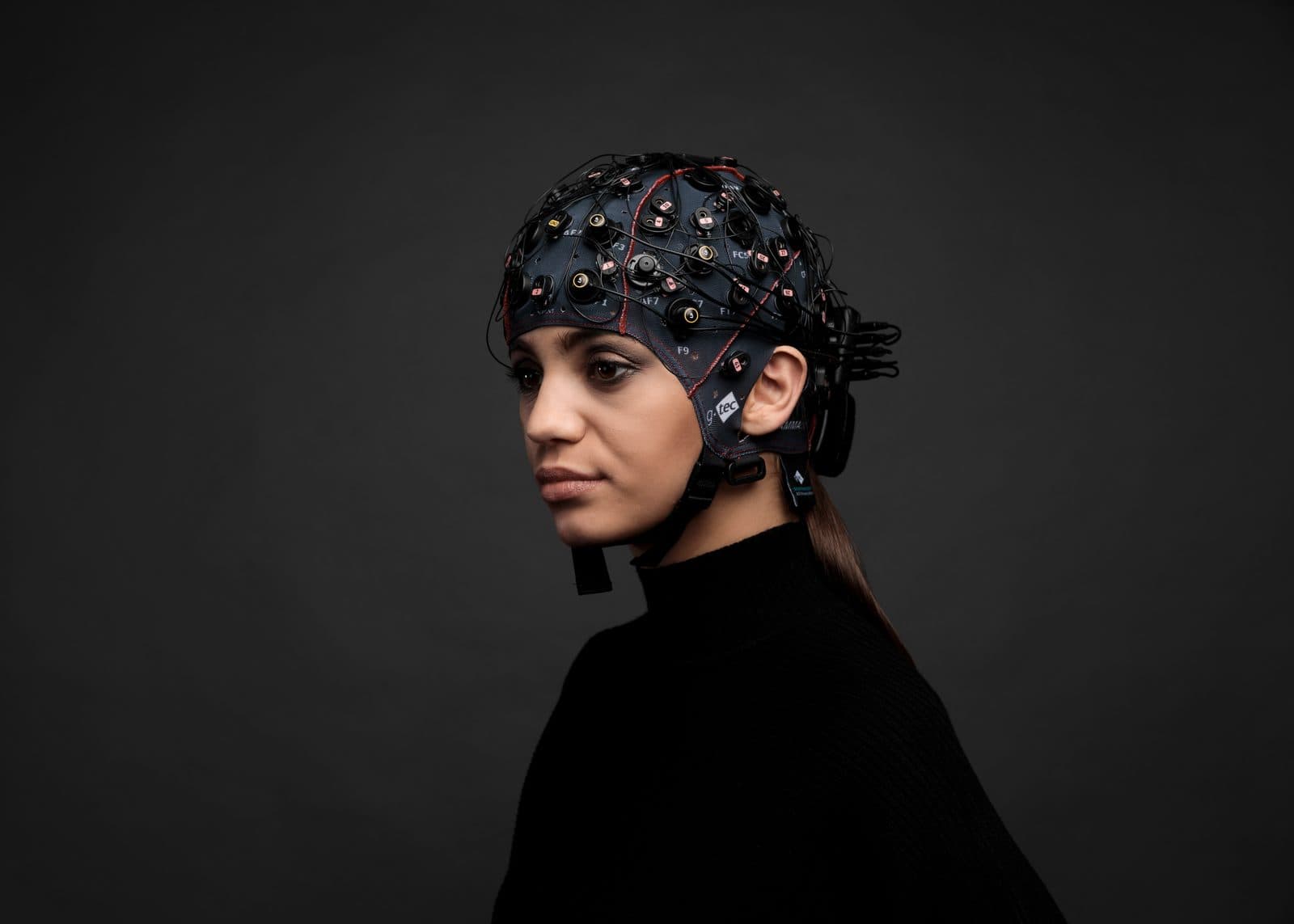
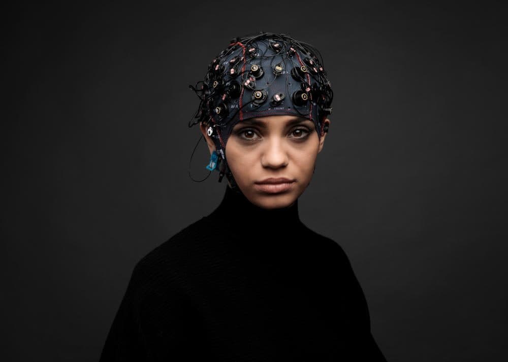
The g.Nautilus fNIRS cap: EEG electrodes and fNIRS optodes

Highlights

  • EEG and fNIRS are acquired at the same sampling rate, synchronised by trigger signals or merged into one data set by g.HIsys Professional
  • LED sources at 760 and 850 nm separate oxyhaemoglobin (HbO) from deoxyhaemoglobin (HbR); continuous-wave (CW) measurement
  • Time-multiplexed scanning illuminates several cortical regions at once
  • Parallel detector readout and adaptive gain switching give a dynamic range of up to 50 dBopt
  • Real-time acquisition, feature extraction and classification in g.HIsys Professional; g.BSanalyze for offline analysis
  • 8 optically isolated TTL inputs for event triggers

Technical specifications

EEG channels
8 / 16 / 32 / 64
EEG sampling rate
250 / 500 Hz (8, 16, 32 channels) · 250 Hz (64 channels)
EEG electrodes
g.SCARABEO active
Cap
g.GAMMAcap: dark blue holders for EEG, light blue for optodes
Sources / detectors
8 or 16 / 8 or 16
Optode holders
32 (on the g.GAMMAcap)
Wavelengths
760 nm and 850 nm
fNIRS sampling rate
70–240 Hz
Detector
Si photodiode or APD · < 1 pW, < 0.1 pW with APD
Dynamic range
50 dBopt
Technique
Continuous wave (CW)
Data transfer
Wi-Fi · USB · internal storage
Event synchronisation
Wireless (LSL) · wired (8-bit TTL)
Battery life
5–6 hours
Software
g.HIsys Professional · g.BSanalyze
Compatible EEG systems
g.Nautilus series · g.HIamp · g.USBamp
EEG and fNIRS

What is measured beneath the skull?

EEG reads the brain's electricity, fNIRS the oxygen in its blood. In one cap they answer the same stimulus on two different time scales. Switch the view, and tap the points in the drawing to see what each layer and part does.

One stimulus, two responses: electrical within milliseconds, haemodynamic within seconds.

Tap the points in the drawing for details. Schematic drawing; layer thicknesses are not to scale.
EEG response · µVStimulus0 200 400 600 msHaemodynamic response · µMStimulus0 5 10 15 20 s
EEG responseHbO, oxyhaemoglobinHbR, deoxyhaemoglobinRaw HbO: heartbeat and Mayer wavesIllustrative signals; time runs faster in the animation.
Absorption (approximate, relative)760850CrossingHbRHbO700750800850900Wavelength, nm

Why two wavelengths?

Oxyhaemoglobin and deoxyhaemoglobin absorb near-infrared light differently: below about 800 nm HbR absorbs more strongly, above it HbO. 760 nm mostly carries the change in HbR, 850 nm the change in HbO. Measuring the attenuation at both wavelengths gives two equations in two unknowns, solved for the concentration change of each haemoglobin.

Change in light attenuation = (HbO's absorption coefficient × change in HbO + HbR's absorption coefficient × change in HbR) × source-detector distance × DPF

The modified Beer-Lambert law, once per wavelength. The DPF (differential pathlength factor) accounts for how many times longer than the distance the light's path through tissue is, because it scatters.

Curves drawn from approximate values in widely used haemoglobin absorption tables (S. Prahl).

Why both

Two modalities that cover each other's gaps

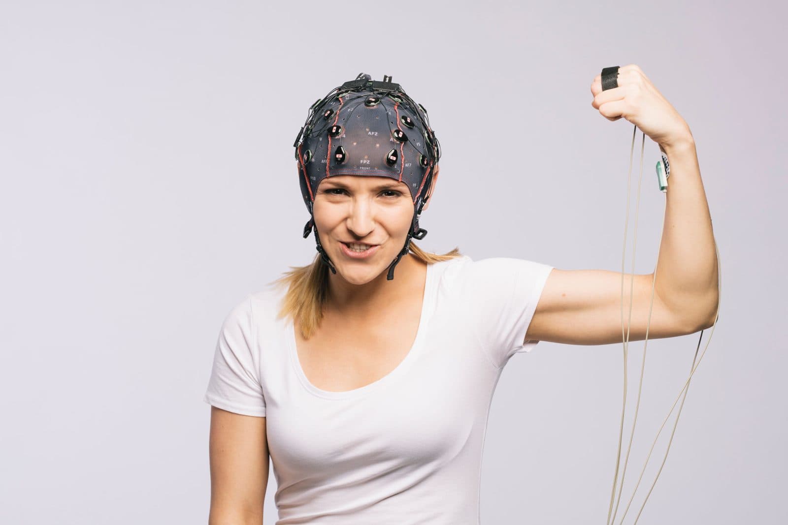
Simultaneous recording with an EEG and fNIRS cap

EEG records the voltage changes of synchronised cortical activity directly, at millisecond resolution; it is the tool for fast dynamics such as motor imagery, P300, SSVEP and ERPs, but its spatial resolution stays at the centimetre scale. fNIRS measures the change in blood flow and oxygenation in active regions with near-infrared light: its response takes seconds, but it maps active cortex to the millimetre. Recording both shows when and where brain activity happens at the same time (Sensors, MDPI 2022; Frontiers in Neuroergonomics 2022; Scientific Reports 2023).

  • Millisecond temporal resolution
  • Millimetre spatial resolution
  • HbO and HbR
Hardware

g.GAMMAcap: both modalities in one cap

g.Nautilus fNIRS on the g.GAMMAcap

The g.GAMMAcap takes up to 64 EEG channels and 32 fNIRS optode holders. Dark blue holders carry 8, 16, 32 or 64 g.SCARABEO active EEG electrodes; light blue holders carry 16 light sources and 16 detectors. Low-power optodes over the frontal cortex follow cognitive load, working memory and executive function; high-power optodes over the sensorimotor cortex follow motor imagery and movement-related activation.

  • 10-20 system positions
  • Frontal optodes
  • Sensorimotor optodes

Applications

g.tec
Contact

Let's plan your project together.

Talk to our team about device selection, protocol design, installation and software integration. We will work out the technical configuration that best fits your research or clinical goal.

Request a quote

Call us +90 554 381 46 20Serving all of Türkiye · reply within 24 hours on working days