Bone defects, fractures and orthopaedic surgical applications.
Illustrative application image
Overview
Different tissue targets, from bone to tendon
Orthopaedics encompasses bone, cartilage, tendons, ligaments and muscle. It shares many tissue targets with sports medicine; the approach depends on the injury, surgical requirements and rehabilitation plan. PRP and platelet gel studies should be assessed separately for each tissue group.
Bone biology
Osteogenesis, osteoinduction and osteoconduction
Bone repair involves bone-forming cells, signals regulating cell differentiation, and a structural scaffold for tissue ingrowth. Platelets carry growth factors including PDGF, TGF-beta, FGF and EGF. This biological role does not establish the same clinical outcome for every PRP preparation or platelet gel system.
Osteogenesis: formation of new bone tissue.
Osteoinduction: signals supporting differentiation into bone-forming cells.
Osteoconduction: a structural scaffold supporting cellular and vascular ingrowth.
Grafts and non-union
Bone grafts, fusion and non-union
Platelet gel may be considered alongside autografts, allografts or synthetic bone grafts in an appropriate surgical context. Bone formation and remodelling are research outcomes in fractures, spinal fusion and non-union. Mechanical stability, circulation and infection must also be considered in non-union treatment planning.
Fracture healing and callus formation
Spinal fusion
Resistant non-union cases
Grafts and tissue engineering
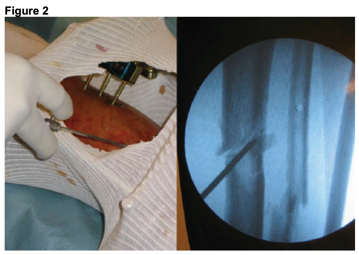
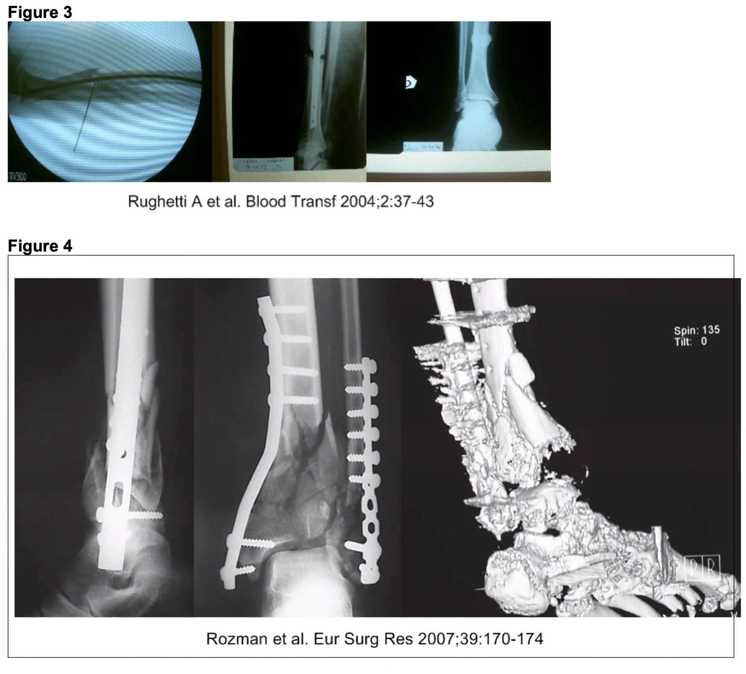
Figure 2. Procedure image and radiological control of an orthopaedic application. Case image supplied by Nevosa; it does not independently establish comparative treatment efficacy.Figures 3–4. Radiological images of bone cases; the lower panel shows fixation hardware and three-dimensional CT assessment. References printed on the figure: Rughetti A et al., Blood Transf, 2004; 2:37–43; Rozman et al., Eur Surg Res, 2007; 39:170–174. Supplied by Nevosa.
Joint replacement
Research outcomes in hip and knee replacement
Platelet gel studies in total hip and knee replacement examine postoperative blood loss, transfusion requirements, pain, osseointegration and hospital stay. Findings depend on preparation, study design and standard care. Patient counts and transfusion comparisons without detailed publication information are not presented as general efficacy claims.
Cartilage
Cartilage defects and marrow stimulation
Cartilage loss and joint degeneration are contexts for research into biological support. Platelet products used with marrow stimulation or surgery may be assessed for pain, function and tissue response. An isolated patient series does not establish the same benefit for every cartilage lesion.
Tendons and ligaments
Tissue structure, loading and repair
Tendons connect muscles to bones; ligaments connect bones to each other. Mechanical loading, collagen organisation and tissue-specific healing capacity matter in injury assessment. Tendinitis and degenerative tendinopathy are distinct. Preparation and application must be considered when selecting PRP or platelet gel; gel and injectable PRP should not be assumed interchangeable.
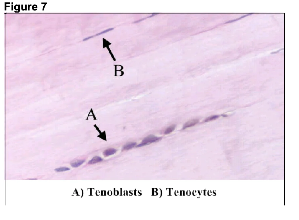
Tenocytes and tenoblasts
Collagen synthesis and matrix organisation
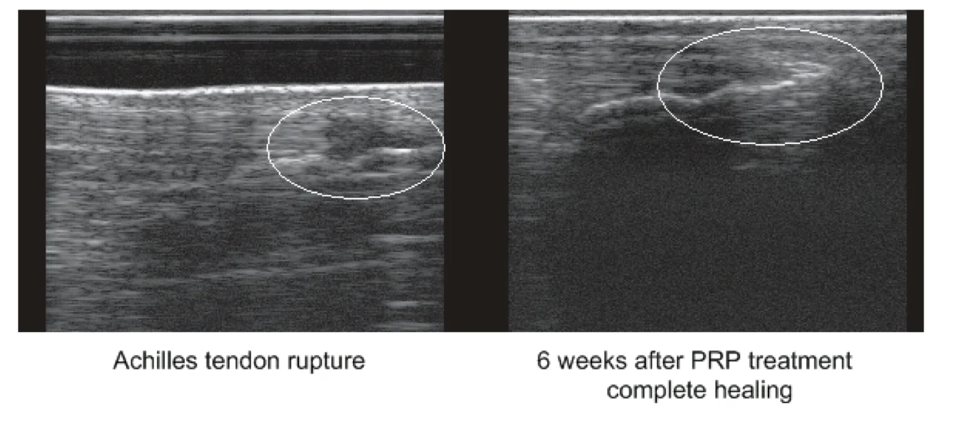
Ultrasound-guided assessment
Rehabilitation and loading plan
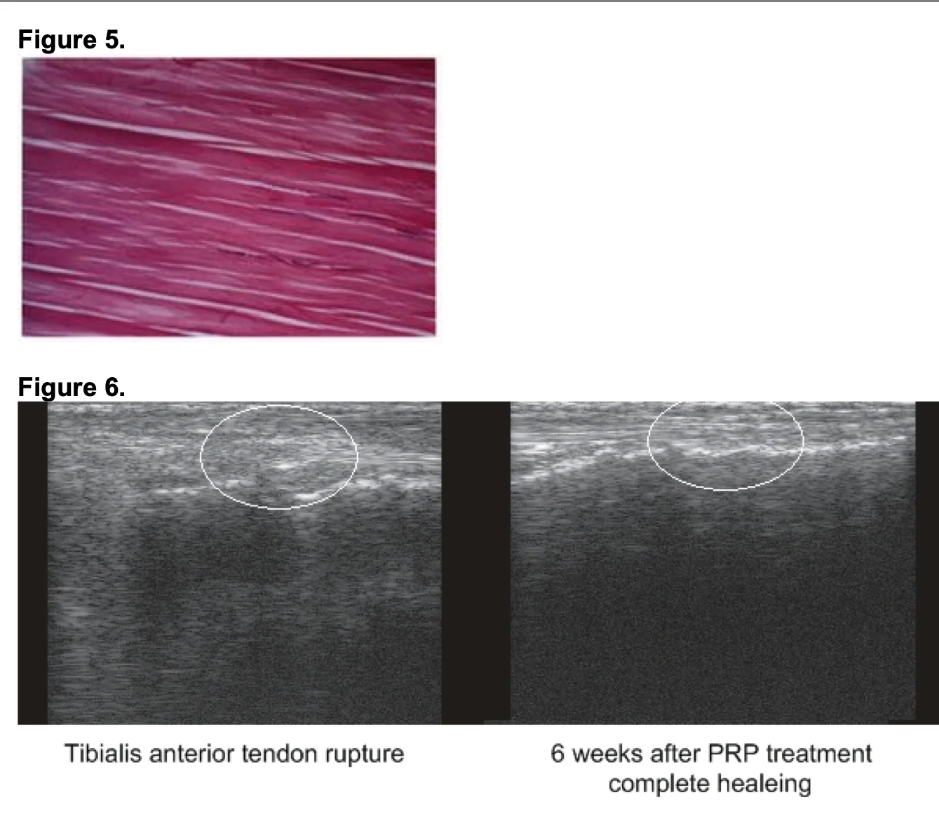
Figures 5–6. Tendon histology above; baseline and six-week ultrasound images of a tibialis anterior tendon lesion following PRP application below. The healing description on the image applies to this case and does not guarantee outcomes or timing. Supplied by Nevosa.Achilles tendon lesion: baseline and six-week ultrasound follow-up after PRP application. The healing reported on the image is an individual case example and should be assessed alongside function and clinical examination. Supplied by Nevosa.Figure 7. Tenoblasts (A) and tenocytes (B) in tendon tissue. This histological figure illustrates cellular structure rather than a before-and-after treatment comparison. Supplied by Nevosa.
Research model
Growth factors and cellular response
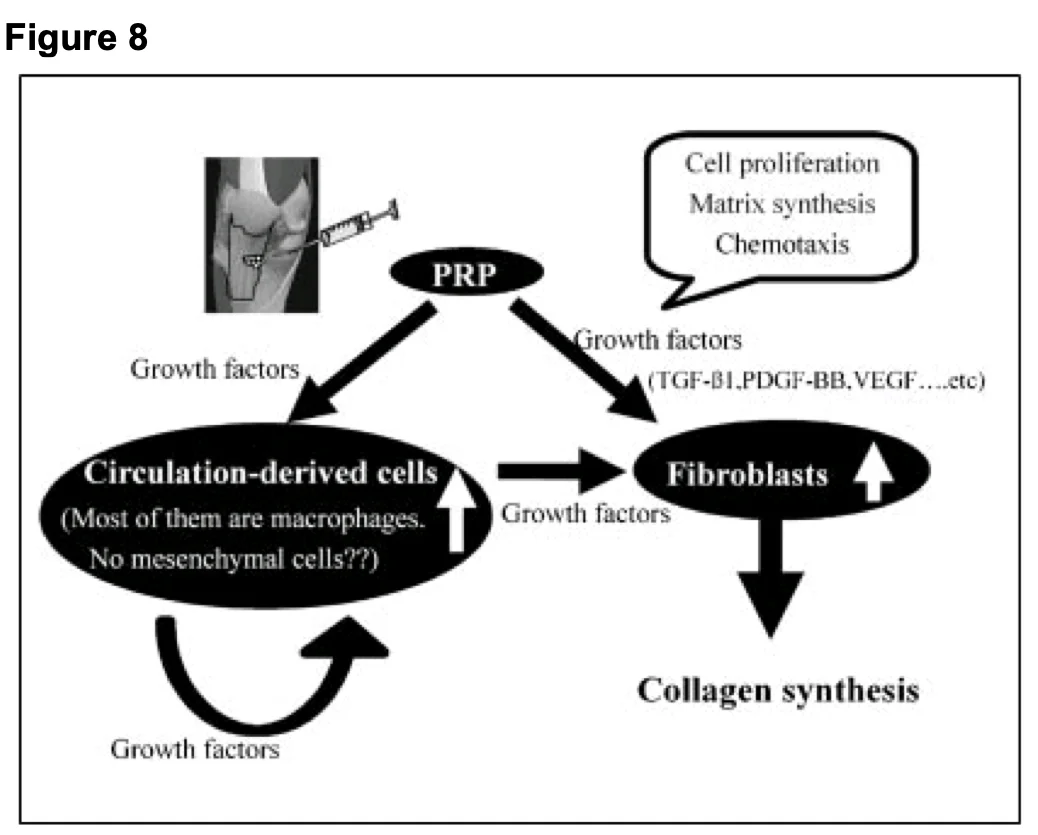
Experimental models investigate platelet-derived growth factors in cell migration, macrophage activity, fibroblast-like cells and collagen synthesis. The supplied text cites Kajikawa and colleagues (Journal of Cellular Physiology, 2008; 215:837–854). An experimental mechanism alone does not validate every clinical application.
Figure 8. Conceptual model linking PRP-derived growth factors, circulating cells and fibroblast responses. It illustrates potential mechanisms related to collagen synthesis, not a measurement of Plateltex-specific clinical efficacy. Supplied by Nevosa.
Surgical repair
Outcomes in ACL and rotator cuff research
Anterior cruciate ligament (ACL) reconstruction and rotator cuff repair are research contexts for platelet products used with surgery. Outcome measures include pain scores, analgesic use, range of motion, function and time to return to activity.
Visual Analogue Scale (VAS)
Analgesic use
Joint range of motion
Function and return to daily activities
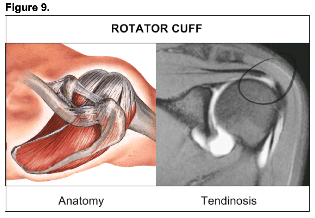
Figure 9. Rotator cuff anatomy on the left and an MRI example labelled tendinosis on the right. This provides anatomical and imaging context rather than a treatment outcome comparison. Supplied by Nevosa.
Regional examples
Ankle, knee, shoulder, elbow and hand
The following are application and research examples discussed in the supplied material. They are not a list of approved Plateltex indications or a treatment recommendation for every condition.
Elbow: lateral epicondylitis and partial distal biceps tears.
Hand: thumb ulnar collateral ligament injuries.
Figure context
Reading schematics and case examples
Figure 1 is a bone–PRP scaffold schematic involving thrombin-mediated platelet activation; it does not directly depict Plateltex’s batroxobin-based gel preparation. Other figures show bone cases, tendon imaging, cellular models and rotator cuff anatomy. Follow-up times and results in case images apply only to the illustrated example.