Tissue repair contexts in dental implants and maxillofacial surgery.
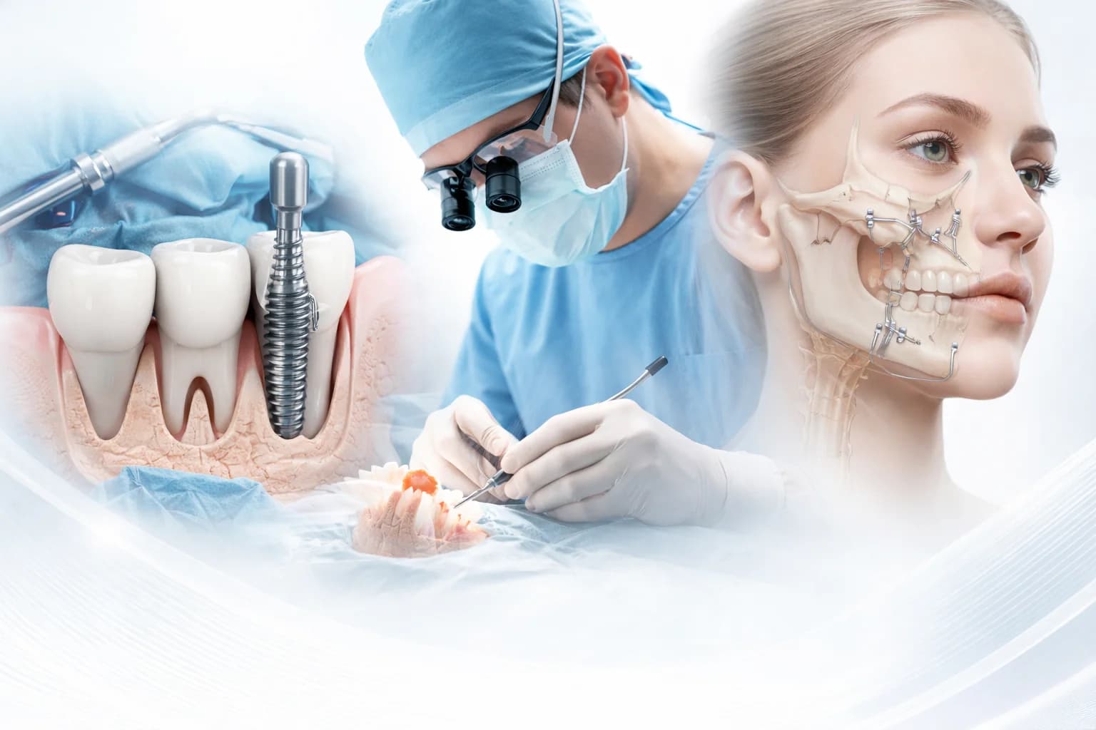
Illustrative application image
Scientific background
PRP and bone grafts: an early study
Robert E. Marx and colleagues’ 1998 study is an early scientific contribution concerning platelet-rich plasma (PRP) used with bone grafts. It belongs to the wider PRP literature; by itself it does not validate the clinical efficacy of the Plateltex system or every batroxobin-prepared gel configuration.
Marx RE et al.
Oral Surg Oral Med Oral Pathol Oral Radiol Endod
1998; 85:638–646
Clinical context
Applications in oral and maxillofacial surgery
The supplied Plateltex material discusses dental implants, alveolar clefts, sinus lift procedures, mandibular and maxillary fractures, and oronasal fistulas as contexts for platelet gel. Selection depends on the defect, graft material and surgical plan.
Dental implants and sinus lift
Alveolar clefts and bone defects
Mandibular and maxillary fractures
Oronasal fistulas
Research outcomes
How is healing assessed?
Research in this area examines healing time, postoperative swelling and pain, soft tissue repair, bone formation and bone density. Findings depend on study design and preparation; the same benefit or faster healing should not be assumed for every patient or procedure.
Healing time
Swelling and pain
Soft tissue repair
Bone regeneration and density
Grafts and biology
Three concepts with different roles
Platelet gel may be considered alongside bone graft materials in an appropriate surgical context. The mechanisms below are distinct; platelet gel alone should not be assumed to replace osteogenic cells or an osteoconductive bone graft.
Osteoinduction: signals supporting differentiation of precursor cells into bone-forming cells.
Osteogenesis: formation of new bone through bone-forming cells.
Osteoconduction: a structural scaffold supporting cellular and vascular ingrowth.
Case and histology
Bone defect after a mandibular cyst
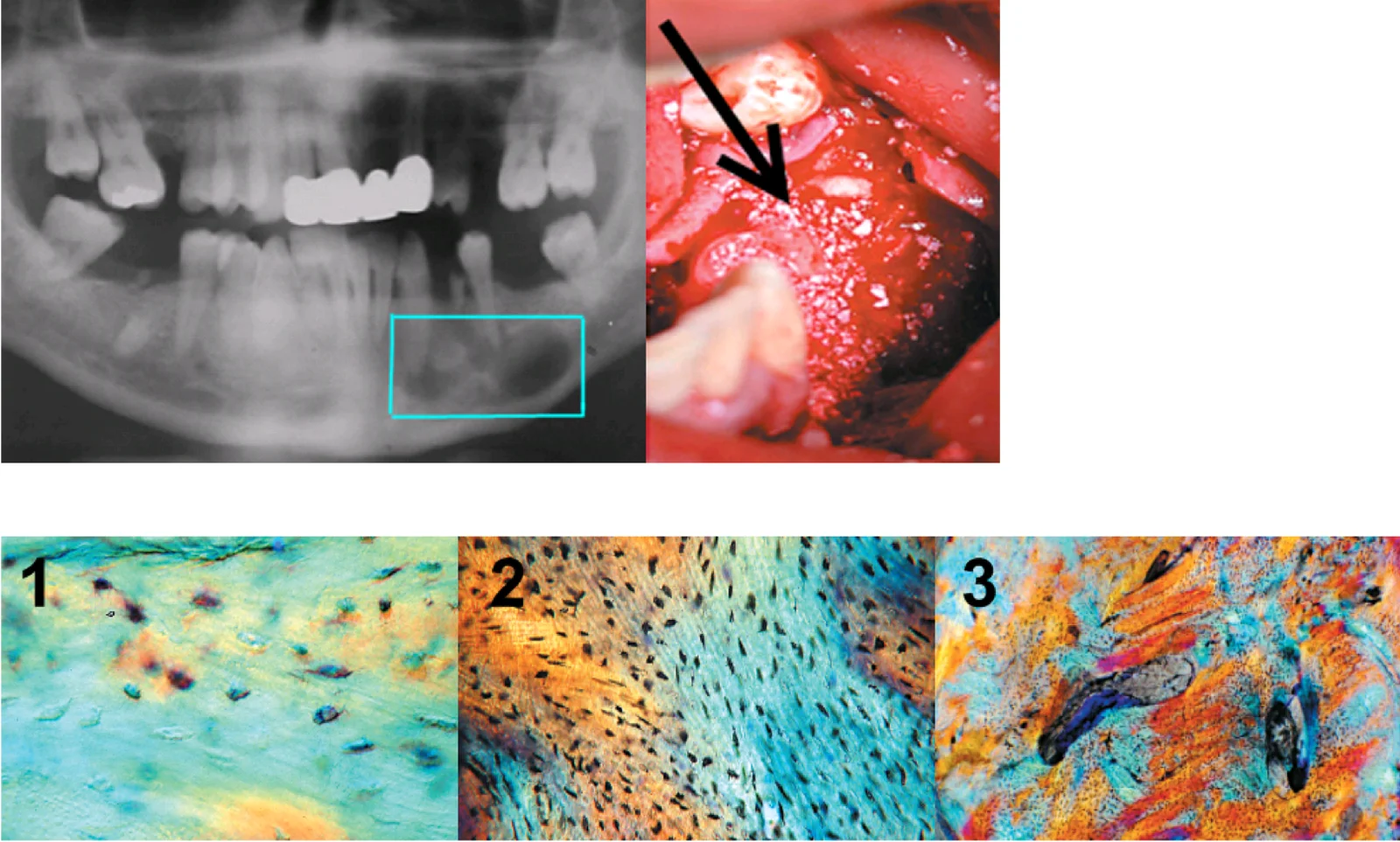
The supplied Pappalardo and colleagues reference describes a mandibular defect filled with particulate bone graft and platelet gel after cyst removal. Its figure description identifies (1) new osteoblast formation around graft particles, (2) low-magnification organisation of new bone and (3) vascular development accompanying bone formation. This is a histological example from a specific case, not general proof of clinical efficacy across all applications.
Pappalardo S et al.
Implantologia Orale
2007; 2:45–49
Case images of a defect following mandibular cyst removal and graft placement. Lower panels: (1) osteoblast formation, (2) organisation of new bone, (3) vascular development. Description and reference supplied by Nevosa: Pappalardo S et al., Implantologia Orale, 2007; 2:45–49.