Wound care contexts including diabetic foot ulcers and pressure injuries.
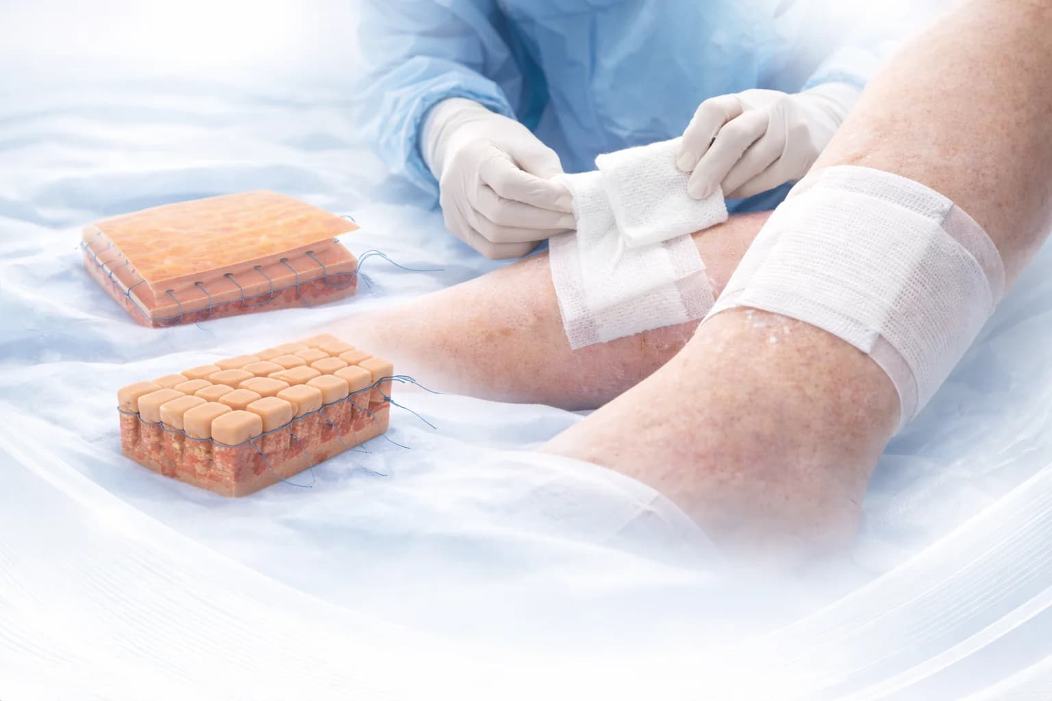
Illustrative application image
Scientific background
Platelet-derived components and wound healing
Knighton and colleagues’ studies in the 1980s are early scientific contributions to understanding platelet-derived components in wound healing. These historical studies concern broader platelet-based approaches and should not be treated as direct evidence for Plateltex across every chronic wound type.
Annals of Surgery, 1982; 196:379–388
Annals of Surgery, 1986; 204:322–330
Clinical context
Which wound types are discussed?
The supplied Plateltex material discusses pressure injuries, diabetic ulcers and diabetic foot wounds, post-traumatic lesions, vascular ulcers and spider-bite lesions. Wound cause, circulation, signs of infection and current care must be assessed before considering a biological approach.
Pressure injuries
Diabetic ulcers and diabetic foot wounds
Post-traumatic lesions
Vascular ulcers
Spider-bite lesions
Care approach
A complementary approach for resistant wounds
Autologous platelet gel may be considered through clinical assessment for wounds that do not heal despite conventional local care and treatment of underlying causes. It does not replace standard wound care or cause-directed treatment. Suitability depends on the product instructions and the patient’s clinical condition.
Multiple factors
Assess the causes behind the wound
Chronic wounds are often associated with multiple factors. Ischaemia, metabolic disorders, bacterial contamination or infection, and nutritional deficiencies can affect healing. A care plan requires appropriate assessment and management of these factors.
Circulation and ischaemia
Metabolic status
Contamination and infection
Nutritional status
Outcome monitoring
Wound closure and changes in size
Changes in wound area, closure and healing time are monitored against baseline measurements. Response varies by wound type, underlying causes and care conditions; a single overall success rate does not represent different patient populations.
Baseline wound size
Change in wound area
Complete closure
Follow-up duration and care conditions
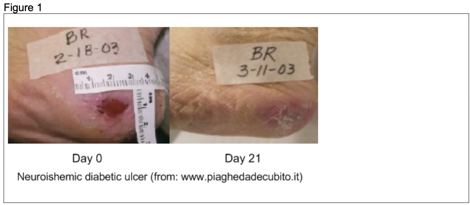
Figure 1. Neuroischaemic diabetic ulcer at baseline (day 0) and day 21. This case does not establish a closure timeline for other wounds. Source printed on the figure: piaghedadecubito.it; supplied by Nevosa.